 Back
Muscles and Muscle Tissue: Structure, Function, and Physiology
Study Guide - Smart Notes
 Tailored notes based on your materials, expanded with key definitions, examples, and context.
Muscle Tissue Types
Overview of Muscle Tissue
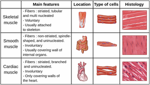
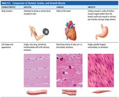
Muscle tissue is specialized for contraction and is essential for movement, posture, and vital functions such as circulation and digestion. There are three main types of muscle tissue: skeletal, cardiac, and smooth muscle.
Skeletal muscle: Voluntary, striated, multinucleated fibers; attached to bones for movement.
Cardiac muscle: Involuntary, striated, branched, and usually uninucleated; forms the walls of the heart.
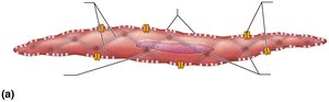
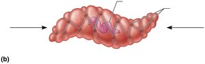
Smooth muscle: Involuntary, non-striated, spindle-shaped, and uninucleated; found in walls of internal organs.
Structure and Organization of Skeletal Muscle
Connective Tissue Sheaths
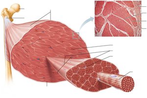
Skeletal muscle is organized into bundles, each surrounded by connective tissue sheaths:
Epimysium: Surrounds the entire muscle.
Perimysium: Surrounds each fascicle (bundle of muscle fibers).
Endomysium: Surrounds individual muscle fibers.
Structural and Organizational Levels
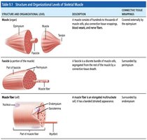
Muscle structure is hierarchical, from the whole muscle down to the myofilaments:
Structure | Description | Connective Tissue Wrapping |
|---|---|---|
Muscle (organ) | Hundreds to thousands of muscle cells, blood vessels, nerve fibers | Epimysium |
Fascicle | Discrete bundle of muscle cells | Perimysium |
Muscle fiber (cell) | Elongated multinucleate cell | Endomysium |
Microscopic Anatomy of Skeletal Muscle
Muscle Fiber Structure
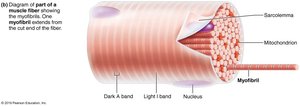
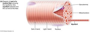
Each muscle fiber contains myofibrils, mitochondria, and is surrounded by the sarcolemma (cell membrane).
Myofibrils: Rod-like structures responsible for contraction.
Sarcolemma: Plasma membrane of the muscle cell.
Mitochondria: Provide energy for contraction.
Striations and Sarcomere Structure
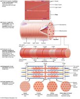
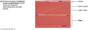
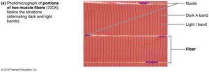
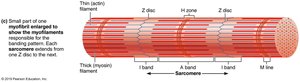
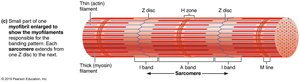
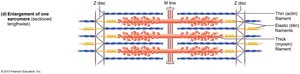
Striations are alternating dark (A bands) and light (I bands) regions, resulting from the arrangement of myofilaments.
A band: Dark region, contains both actin and myosin.
I band: Light region, contains only actin.
H zone: Lighter region in the middle of the A band.
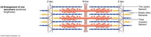
M line: Center of the sarcomere, anchors myosin filaments.
Z disc: Boundary of the sarcomere, anchors actin filaments.
Myofilaments: Actin and Myosin
Myofilaments are the contractile proteins within the sarcomere:
Actin (thin filament): Anchored to Z discs, contains myosin-binding sites.
Myosin (thick filament): Anchored at M line, has heads that bind to actin and hydrolyze ATP.
Clinical Application: Duchenne Muscular Dystrophy
Pathophysiology
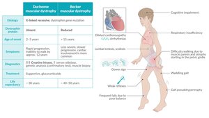
Duchenne muscular dystrophy (DMD) is a severe hereditary neuromuscular disease caused by mutations in the dystrophin gene, leading to progressive muscle degeneration and weakness.
Symptoms: Muscle weakness, loss of ambulation, respiratory insufficiency.
Diagnosis: Genetic testing, elevated creatine kinase.
Excitation-Contraction Coupling
Neuromuscular Junction
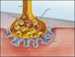
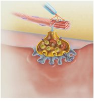
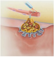
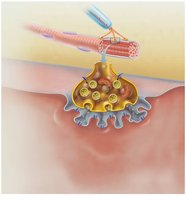
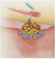
The neuromuscular junction (NMJ) is the site where a motor neuron communicates with a muscle fiber:
Axon terminal: Releases acetylcholine (ACh) into the synaptic cleft.
Junctional folds: Increase surface area for ACh receptors.
Synaptic vesicles: Contain ACh.
Steps of Muscle Fiber Excitation
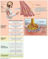
The process of muscle contraction involves several steps:
Action potential arrives at axon terminal.
Voltage-gated Ca2+ channels open; Ca2+ enters axon terminal.
Ca2+ triggers exocytosis of ACh.
ACh binds to receptors on sarcolemma, opening ion channels.
Na+ enters, K+ exits, generating end plate potential.
ACh is degraded by acetylcholinesterase, terminating the signal.
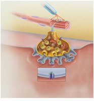
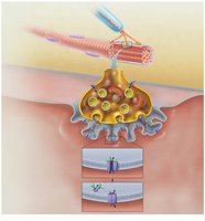
Action Potential and Muscle Contraction
The resting sarcolemma is polarized, and muscle contraction is initiated by changes in membrane potential:
Depolarization: Na+ channels open, Na+ enters.
Repolarization: K+ channels open, K+ exits.
Cross Bridge Cycle
Mechanism of Contraction
The cross bridge cycle describes how myosin heads pull actin filaments toward the center of the sarcomere:
Cross bridge formation: Myosin head binds to actin.
Power stroke: Myosin head pivots, pulling actin toward M line; ADP and Pi are released.
Cross bridge detachment: ATP binds to myosin, causing detachment from actin.
Cocking of myosin head: ATP is hydrolyzed, re-energizing the myosin head.
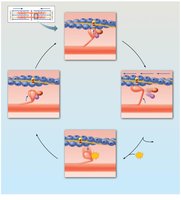
Motor Units and Muscle Twitch
Motor Unit Structure
A motor unit consists of a motor neuron and all the muscle fibers it innervates. The number of fibers per motor unit varies by muscle function. 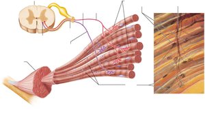
Muscle Twitch Phases
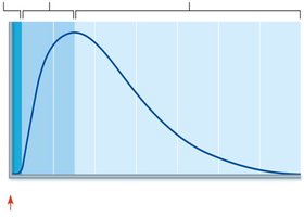
A muscle twitch is a single contraction event, consisting of three phases:
Latent period: Excitation-contraction coupling; no tension.
Contraction period: Cross bridge formation; tension increases.
Relaxation period: Ca2+ reentry into SR; tension decreases.
Summation and Tetanus
Temporal Summation
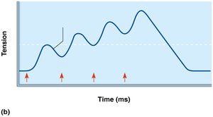
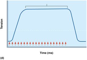
Repeated stimuli before relaxation is complete lead to increased tension (summation), and at high frequency, fused tetanus occurs.
Unfused tetanus: Partial relaxation between stimuli.
Fused tetanus: No relaxation; maximal tension.
Muscle Contraction Types
Isotonic and Isometric Contractions
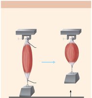
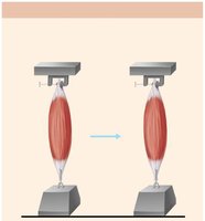
Isotonic contraction: Muscle shortens as it contracts, tension remains constant.
Isometric contraction: Muscle does not shorten, tension increases.
Energy for Muscle Contraction
ATP Regeneration Pathways
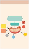
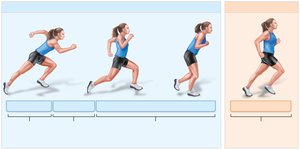
Muscle fibers regenerate ATP through three main pathways:
Direct phosphorylation: Creatine phosphate + ADP → ATP (short duration).
Anaerobic glycolysis: Glucose → ATP + lactic acid (moderate duration).
Aerobic respiration: Glucose, fatty acids, amino acids → ATP + CO2 + H2O (long duration).
Factors Affecting Muscle Contraction
Force, Velocity, and Duration
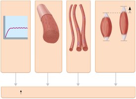
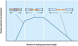
Force: Increased by high frequency stimulation, large number of fibers recruited, large fibers, optimal sarcomere length.
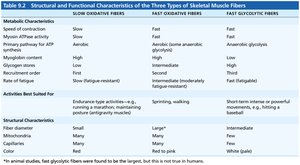
Velocity and duration: Influenced by fiber type, load, and recruitment.
Smooth Muscle Structure and Function
Innervation and Contraction
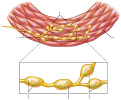
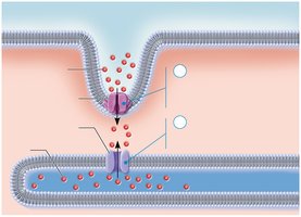
Smooth muscle is innervated by autonomic nerve fibers, and contraction is regulated by Ca2+ entry and calmodulin activation.
Varicosities: Release neurotransmitters into diffuse junctions.
Caveolae: Membrane infoldings for Ca2+ entry.
Dense bodies: Anchor actin filaments.
Comparison of Muscle Types
Structural and Functional Differences
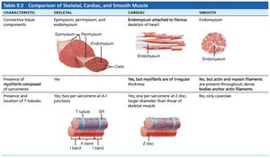
Feature | Skeletal | Cardiac | Smooth |
|---|---|---|---|
Striations | Yes | Yes | No |
Control | Voluntary | Involuntary | Involuntary |
Cell shape | Long, cylindrical | Branched | Spindle-shaped |
Nuclei | Multinucleated | Uninucleated | Uninucleated |
Location | Attached to bones | Heart | Walls of organs |
Muscle Development, Regeneration, and Aging
Muscle Development
Skeletal muscle fibers form by fusion of myoblasts during embryonic development.
Myoblasts: Embryonic cells that fuse to form multinucleate muscle fibers.
Regeneration and Aging
Regeneration: Mature skeletal muscle fibers cannot undergo mitosis; repair is limited.
Aging: Muscle tissue is replaced by connective and adipose tissue, leading to decreased strength and flexibility.
Additional info: This guide expands on brief points with academic context, definitions, and examples to ensure completeness and clarity for exam preparation.
