 Back
Muscles and Muscle Tissue: Structure, Function, and Contraction Mechanisms
Study Guide - Smart Notes
 Tailored notes based on your materials, expanded with key definitions, examples, and context.
Muscle Tissue Types
Overview of Muscle Tissue
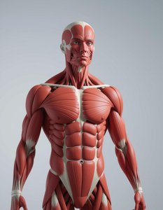
Muscle tissue is essential for movement, stability, and various physiological functions. There are three basic types of muscle tissue in the human body: skeletal, cardiac, and smooth muscle. Each type has distinct structural and functional characteristics.
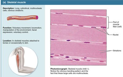
Skeletal Muscle: Responsible for voluntary movement, attached to bones.
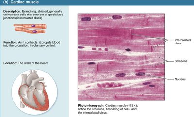
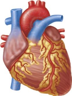
Cardiac Muscle: Found only in the heart, responsible for pumping blood, involuntary.
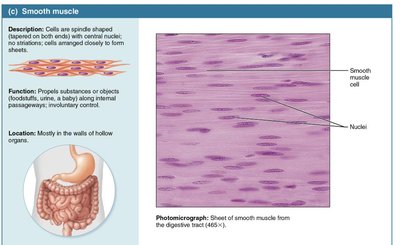
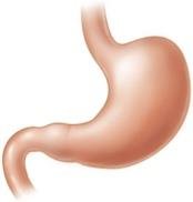
Smooth Muscle: Found in the walls of internal organs, responsible for involuntary movements.
Comparison of Muscle Tissue Types
Characteristic | Skeletal | Cardiac | Smooth |
|---|---|---|---|
Body Location | Attached to bones or skin | Walls of the heart | Walls of hollow organs |
Cell Shape & Appearance | Long, cylindrical, multinucleate, striated | Branching chains, uni- or binucleate, striated | Spindle-shaped, uninucleate, no striations |
Control | Voluntary | Involuntary | Involuntary |
Function | Movement, posture, heat | Pumping blood | Propelling substances |
Characteristics and Functions of Muscle Tissue
Key Properties
Muscle tissue exhibits four main characteristics:
Excitability: Ability to respond to stimuli by changing membrane potential.
Contractility: Ability to shorten forcibly when stimulated.
Extensibility: Ability to stretch.
Elasticity: Ability to recoil after stretching.
Functions
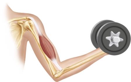
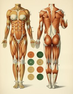
Movement: Skeletal muscles contract to produce body movements.
Maintain Posture: Muscles stabilize joints and maintain posture.
Stabilizing Joints: Muscle tendons reinforce and stabilize joints.
Heat Generation: Muscle contraction generates heat, maintaining body temperature.
Development and Structure of Skeletal Muscle
Developmental Aspects
All muscle tissues develop from myoblasts, which are embryonic mesoderm cells. Multinucleated skeletal muscle cells form by fusion of many myoblasts, while cardiac and smooth muscle myoblasts do not fuse but develop gap junctions. 
Gross Structure of Skeletal Muscle
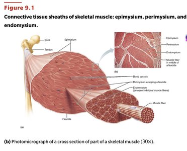
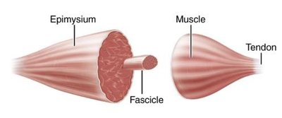
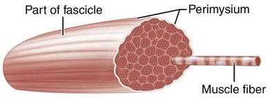
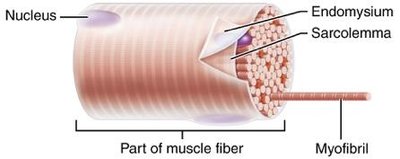
Skeletal muscle is organized into layers of connective tissue:
Epimysium: Surrounds the entire muscle.
Perimysium: Surrounds each fascicle (bundle of muscle fibers).
Endomysium: Surrounds each muscle fiber.
Microscopic Structure of Skeletal Muscle
Myofibrils and Sarcomeres
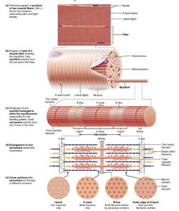
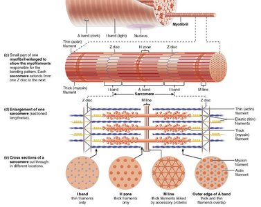
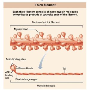
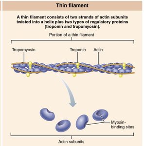
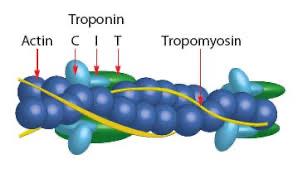
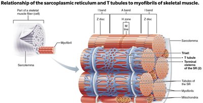
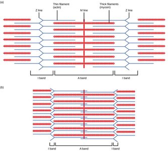
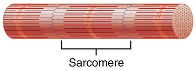
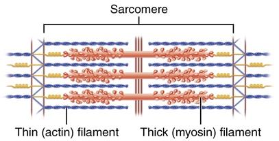
Myofibrils are contractile units within muscle fibers, composed of repeating sarcomeres. Sarcomeres are the functional units, containing actin (thin) and myosin (thick) filaments.
Z-discs: Boundaries of the sarcomere.
A band: Dark band, contains thick filaments.
I band: Light band, contains thin filaments.
Sarcoplasmic Reticulum and T Tubules
Sarcoplasmic Reticulum (SR)
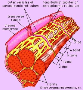
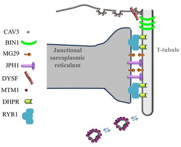
The SR is a network of tubules surrounding each myofibril, storing calcium ions (Ca2+) essential for muscle contraction. Upon stimulation, the SR releases Ca2+ into the sarcoplasm, triggering contraction. 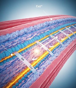
T Tubules
T tubules are invaginations of the sarcolemma that transmit action potentials deep into the muscle fiber, ensuring coordinated contraction. They are closely associated with the SR, forming triads that facilitate Ca2+ release. 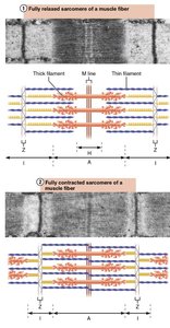
Sliding Filament Model of Muscle Contraction
Mechanism
The sliding filament model states that during contraction, thin filaments (actin) slide past thick filaments (myosin), increasing their overlap and shortening the sarcomere. This process requires ATP and Ca2+.
Myosin heads bind to actin, forming cross-bridges.
Power stroke pulls actin toward the M line.
ATP binding causes myosin to detach from actin.
ATP hydrolysis re-cocks the myosin head.
Neuromuscular Junction and Excitation-Contraction Coupling
Neuromuscular Junction (NMJ)
The NMJ is the synapse between a motor neuron and a muscle fiber. When a motor neuron fires, it releases acetylcholine (ACh), which binds to receptors on the sarcolemma, depolarizing the muscle fiber and initiating contraction.
Acetylcholinesterase (AChE): Breaks down ACh, preventing continuous stimulation.
Excitation-Contraction Coupling
This sequence links the action potential in the sarcolemma to muscle contraction:
Action potential travels along sarcolemma and down T tubules.
Triggers Ca2+ release from SR.
Ca2+ binds to troponin, exposing myosin-binding sites on actin.
Myosin binds to actin, initiating cross-bridge cycle.
Motor Units and Muscle Twitch
Motor Unit
A motor unit consists of a motor neuron and all the muscle fibers it innervates. The size of a motor unit varies depending on the muscle's function.
Large motor units: Weight-bearing muscles (e.g., hip).
Small motor units: Muscles requiring fine control.
Muscle Twitch
A muscle twitch is the response of a muscle to a single, brief stimulus. It consists of three phases:
Latent Period: Time between stimulation and start of contraction.
Contraction Phase: Muscle contracts, cross-bridges are active.
Relaxation Phase: Muscle relaxes, Ca2+ is pumped back into SR.
Types of Muscle Contractions
Isometric vs. Isotonic Contractions
Isometric Contraction: Muscle length remains constant, tension increases. Example: Pushing against a wall.
Isotonic Contraction: Muscle tension remains constant, muscle length changes. Example: Lifting a weight.
Concentric: Muscle shortens and does work.
Eccentric: Muscle lengthens while generating force.
ATP Regeneration During Muscle Contraction
Energy Pathways
Muscle contraction requires ATP, which is regenerated by three main pathways:
Direct Phosphorylation: Creatine phosphate donates a phosphate to ADP, forming ATP. Fastest method.
Anaerobic Glycolysis: Glucose is broken down to produce ATP and lactic acid. Does not use oxygen.
Aerobic Respiration: Glucose, fatty acids, and amino acids are broken down in the presence of oxygen to produce ATP. Most efficient, used during prolonged exercise.
Muscle Fatigue and EPOC
Definitions and Causes
EPOC (Excess Post-exercise Oxygen Consumption): Extra oxygen needed after exercise to restore energy reserves and remove lactic acid.
Muscle Fatigue: Decline in muscle force and endurance due to ATP depletion, lactic acid accumulation, and ion imbalances.
Types of Skeletal Muscle Fibers
Classification
Slow Oxidative Fibers (Type I): High endurance, fatigue-resistant.
Fast Oxidative Fibers (Type IIa): Moderate endurance, fatigue-resistant.
Fast Glycolytic Fibers (Type IIb/x): Low endurance, easily fatigued.
Effects of Exercise on Skeletal Muscles
Aerobic vs. Resistance Exercise
Aerobic Exercise: Increases endurance, cardiovascular health, capillary density, and mitochondrial number.
Resistance Exercise: Increases muscle strength and size (hypertrophy), stimulates synthesis of actin and myosin, increases muscle fiber diameter.
Smooth Muscle: Gross and Microscopic Anatomy
Gross Anatomy
Smooth muscle cells are spindle-shaped, uninucleated, and organized into sheets or layers. They lack striations due to the arrangement of actin and myosin filaments.
Microscopic Anatomy
Actin and Myosin Filaments: Present, but not arranged in sarcomeres.
Dense Bodies: Anchor points for actin filaments.
Intermediate Filaments: Provide structural support.
Lack of T Tubules: Calcium influx facilitated by caveolae.
Sarcoplasmic Reticulum: Less developed than in skeletal muscle.
Contractile Mechanisms: Skeletal vs. Smooth Muscle
Comparison
Feature | Skeletal Muscle | Smooth Muscle |
|---|---|---|
Activation | Neuromuscular junction, action potentials | Neurotransmitters, hormones, local factors |
Calcium Source | Sarcoplasmic reticulum | Extracellular fluid and SR |
Regulation | Troponin-tropomyosin complex | Calmodulin and MLCK |
Contraction Speed | Fast | Slow and sustained |
Unitary vs. Multi-Unit Smooth Muscle
Structural and Functional Differences
Unitary Smooth Muscle: Found in walls of hollow organs, electrically coupled by gap junctions, contracts as a unit, responds to various chemical stimuli.
Multi-Unit Smooth Muscle: Cells not connected by gap junctions, each contracts independently, allows for fine motor control, found in iris of eye and arrector pili muscles.
Summary Tables
Comparison of Skeletal, Cardiac, and Smooth Muscle
Characteristic | Skeletal | Cardiac | Smooth |
|---|---|---|---|
Connective Tissue Components | Epimysium, perimysium, endomysium | Endomysium attached to fibrous skeleton | Endomysium |
Presence of Myofibrils/Sarcomeres | Yes | Yes, irregular thickness | No, but actin and myosin present |
Presence and Location of T Tubules | Yes, two per sarcomere at A-I junctions | Yes, one per sarcomere at Z disc | No, only caveolae |
Source of Ca2+ for Contraction | Sarcoplasmic reticulum | SR and extracellular fluid | Extracellular fluid and SR |
Site of Calcium Regulation | Troponin on actin | Troponin on actin | Calmodulin in cytosol |
Speed of Contraction | Slow to fast | Slow | Very slow |
Summary of Key Concepts
Muscle Tissue Types: Skeletal (voluntary, striated), cardiac (involuntary, striated), smooth (involuntary, non-striated).
Functions: Movement, posture, heat generation, joint stabilization.
Skeletal Muscle Structure: Muscle fibers, myofibrils, sarcomeres, connective tissues.
Contraction Mechanisms: Sliding filament model, cross bridge cycle, calcium's role.
Muscle Fiber Types: Slow oxidative, fast oxidative, fast glycolytic.
Muscle Fatigue and EPOC: Causes and recovery mechanisms.
