Back
Muscles and Muscle Tissue: Structure, Function, and Physiology
Study Guide - Smart Notes
 Tailored notes based on your materials, expanded with key definitions, examples, and context.
Muscle Tissue Overview
Introduction to Muscle Tissue
Muscle tissue comprises nearly half of the body's mass and is essential for movement, posture, and heat generation. Muscles convert chemical energy (ATP) into mechanical energy, enabling forceful contractions.
Prefixes: myo-, mys-, and sarco- refer to muscle (e.g., sarcoplasm is muscle cell cytoplasm).
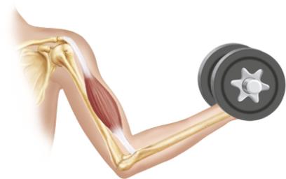
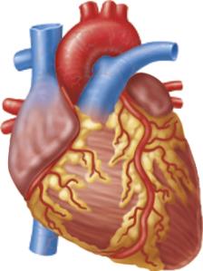
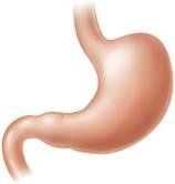
Three types of muscle tissue: Skeletal, Cardiac, Smooth.
Muscle fibers: Elongated muscle cells found in skeletal and smooth muscle, but not cardiac muscle.
Types of Muscle Tissue
Characteristic | Skeletal | Cardiac | Smooth |
|---|---|---|---|
Body Location | Attached to bones or skin | Walls of the heart | Walls of hollow organs (except heart) |
Cell Shape & Appearance | Long, cylindrical, multinucleate, striated | Branching chains, uni- or binucleate, striated | Spindle-shaped, uninucleate, non-striated |
Control | Voluntary | Involuntary | Involuntary |
Characteristics of Muscle Tissue
Excitability (Responsiveness): Ability to receive and respond to stimuli.
Contractility: Ability to shorten forcibly when stimulated.
Extensibility: Ability to be stretched.
Elasticity: Ability to recoil to resting length.
Functions of Muscle Tissue
Produce movement: Locomotion and manipulation (e.g., walking, digestion, pumping blood).
Maintain posture and body position.
Stabilize joints.
Generate heat: As muscles contract.
Skeletal Muscle Structure
Organization and Connective Tissue Sheaths
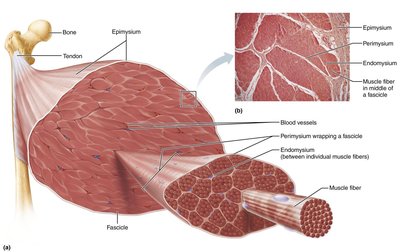
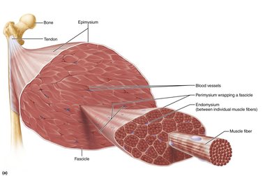
Skeletal muscle is an organ composed of muscle fibers, nerves, blood vessels, and connective tissue sheaths. These sheaths support and reinforce the muscle.
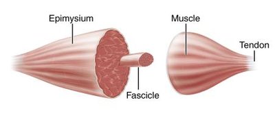
Epimysium: Dense irregular connective tissue surrounding the entire muscle.
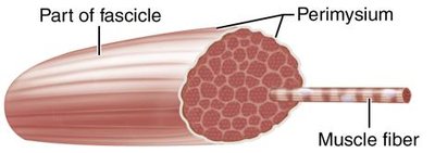
Perimysium: Fibrous connective tissue surrounding fascicles (bundles of muscle fibers).
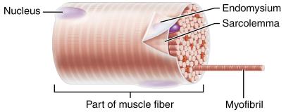
Endomysium: Fine areolar connective tissue surrounding each muscle fiber.
Muscle Attachments
Direct (fleshy) attachment: Epimysium fused to periosteum of bone or perichondrium of cartilage.
Indirect attachment: Connective tissue wrappings extend beyond muscle as a tendon or aponeurosis.
Structural Organization
Level | Description | Connective Tissue |
|---|---|---|
Muscle (organ) | Hundreds to thousands of muscle cells, blood vessels, nerves | Epimysium |
Fascicle | Bundle of muscle cells | Perimysium |
Muscle fiber (cell) | Elongated, multinucleate, striated cell | Endomysium |
Skeletal Muscle Fiber Microanatomy
Microscopic Anatomy
Skeletal muscle fibers are long, cylindrical cells with multiple nuclei. The sarcolemma is the plasma membrane, and the sarcoplasm is the cytoplasm, containing glycosomes (glycogen storage) and myoglobin (O2 storage).
Myofibrils: Densely packed, rodlike elements responsible for muscle contraction and striations.
Sarcoplasmic reticulum (SR): Smooth ER that stores and releases Ca2+.
T tubules: Invaginations of the sarcolemma that conduct impulses deep into the muscle fiber.
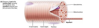
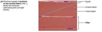
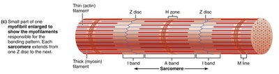
Sarcomere and Myofilaments
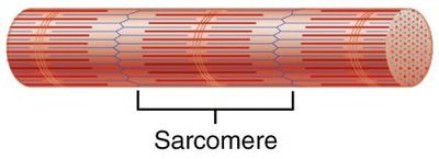
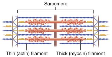
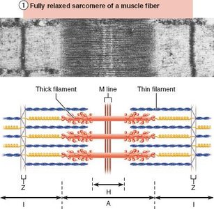
The sarcomere is the smallest contractile unit of muscle, defined as the region between two Z discs. It contains thick (myosin) and thin (actin) filaments arranged in a precise pattern.
A bands: Dark regions containing thick filaments.
I bands: Light regions containing thin filaments.
H zone: Lighter region in the middle of the A band.
M line: Line of protein that bisects the H zone.
Z disc: Sheet of proteins that anchors thin filaments.
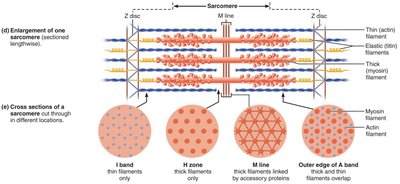
Molecular Composition of Myofilaments
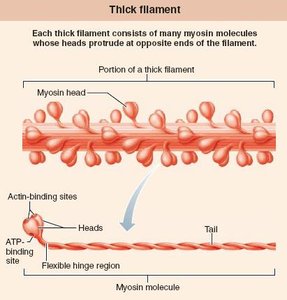
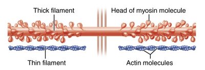
Thick filaments: Composed of myosin molecules with heads that form cross bridges during contraction.
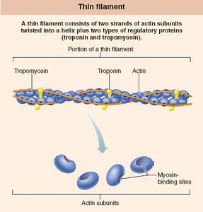
Thin filaments: Composed of actin, tropomyosin, and troponin. Actin has binding sites for myosin heads.
Elastic filaments: Composed of titin, which helps maintain sarcomere structure and elasticity.
Dystrophin: Links thin filaments to the sarcolemma.
Muscle Contraction Physiology
Sliding Filament Model of Contraction
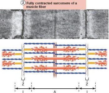
Muscle contraction occurs when thin filaments slide past thick filaments, increasing their overlap. This process is powered by cross bridge cycling between actin and myosin.
During contraction: I bands shorten, Z discs move closer, H zones disappear, and A bands move closer together.
Neuromuscular Junction and Muscle Fiber Excitation
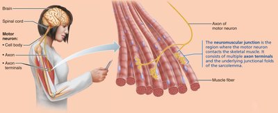
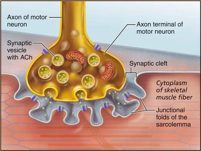
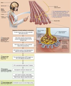
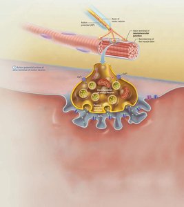
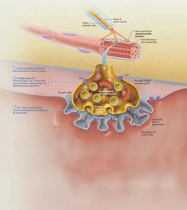
The neuromuscular junction (NMJ) is the site where a motor neuron stimulates a muscle fiber. The neurotransmitter acetylcholine (ACh) is released, triggering an action potential in the muscle fiber.
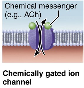
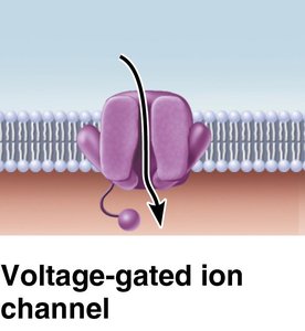
Ion channels: Chemically gated (ACh receptors) and voltage-gated channels are essential for action potential propagation.
Generation of an Action Potential
End plate potential: ACh binding opens ion channels, causing Na+ influx and local depolarization.
Depolarization: If threshold is reached, voltage-gated Na+ channels open, generating an action potential.
Repolarization: Na+ channels close, K+ channels open, restoring resting membrane potential.
Excitation-Contraction (E-C) Coupling
E-C coupling links the action potential to muscle contraction. The AP travels along the sarcolemma and T tubules, triggering Ca2+ release from the SR, which initiates contraction.
Cross Bridge Cycle
Cross bridge formation: Myosin head attaches to actin.
Power stroke: Myosin head pivots, pulling actin filament toward M line.
Cross bridge detachment: ATP binds to myosin, causing detachment.
Cocking of myosin head: ATP hydrolysis re-energizes the myosin head.
Whole Muscle Contraction
Motor Units and Muscle Twitch
Motor unit: A motor neuron and all the muscle fibers it innervates.
Muscle twitch: Response of a muscle to a single stimulus, consisting of latent, contraction, and relaxation phases.
Graded Muscle Contractions
Temporal (wave) summation: Increased frequency of stimulation increases contraction strength.
Recruitment: Increasing stimulus strength recruits more motor units for stronger contractions.
Isotonic and Isometric Contractions
Isotonic contraction: Muscle changes length and moves a load (concentric: shortens; eccentric: lengthens).
Isometric contraction: Muscle tension increases but does not exceed load; muscle does not change length.
Energy for Muscle Contraction
ATP Regeneration Pathways
Direct phosphorylation: Creatine phosphate donates phosphate to ADP to form ATP.
Anaerobic pathway: Glycolysis and lactic acid formation (no oxygen required).
Aerobic pathway: Glycolysis followed by aerobic respiration in mitochondria (requires oxygen).
Equation for direct phosphorylation:
Muscle Fatigue and Recovery
Muscle fatigue: Inability to contract despite stimulation, often due to ionic imbalances, increased inorganic phosphate, or decreased ATP.
Excess postexercise oxygen consumption (EPOC): Oxygen required to restore muscle to resting state.
Factors Affecting Muscle Contraction
Force, Velocity, and Duration
Force: Depends on frequency of stimulation, number of fibers recruited, size of fibers, and degree of stretch.
Velocity and duration: Influenced by muscle fiber type, load, and recruitment.
Fiber Type | Speed | ATP Pathway | Fatigue Resistance | Best Suited For |
|---|---|---|---|---|
Slow Oxidative | Slow | Aerobic | High | Endurance (posture, marathon) |
Fast Oxidative | Fast | Aerobic (some anaerobic) | Intermediate | Sprinting, walking |
Fast Glycolytic | Fast | Anaerobic glycolysis | Low | Short-term intense movements |
Smooth Muscle
Structure and Function
Found in walls of hollow organs (except heart).
Spindle-shaped, uninucleate, non-striated cells.
Organized in longitudinal and circular layers for peristalsis.
Innervated by autonomic nervous system; uses varicosities instead of neuromuscular junctions.
Contraction is slow, sustained, and energy-efficient.
Comparison Table: Skeletal, Cardiac, and Smooth Muscle
Characteristic | Skeletal | Cardiac | Smooth |
|---|---|---|---|
Control | Voluntary | Involuntary | Involuntary |
Striations | Yes | Yes | No |
Cell Shape | Long, cylindrical | Branched | Spindle-shaped |
Connective Tissue | Epimysium, perimysium, endomysium | Endomysium | Endomysium |
Calcium Regulation | Troponin | Troponin | Calmodulin |
Contraction Speed | Slow to fast | Slow | Very slow |
Development and Clinical Aspects
Muscle Development and Regeneration
All muscle tissues develop from embryonic mesoderm cells called myoblasts.
Skeletal muscle fibers form by fusion of myoblasts; cardiac and smooth muscle do not fuse but develop gap junctions.
Regeneration: Skeletal muscle has limited regenerative ability; smooth muscle regenerates throughout life.
Clinical Conditions
Duchenne muscular dystrophy (DMD): X-linked disorder causing muscle degeneration due to defective dystrophin.
Myasthenia gravis: Autoimmune disease where ACh receptors are destroyed, leading to muscle weakness.
Rigor mortis: Postmortem muscle stiffening due to lack of ATP for cross bridge detachment.
Disuse atrophy: Muscle wasting due to inactivity or loss of neural stimulation.