 Back
Muscles and Muscle Tissue: Structure, Function, and Physiology
Study Guide - Smart Notes
 Tailored notes based on your materials, expanded with key definitions, examples, and context.
Muscle Tissue Overview
Types of Muscle Tissue
Muscle tissue is specialized for contraction and is responsible for movement in the body. There are three main types of muscle tissue, each with distinct characteristics and functions:
Skeletal Muscle: Attached to bones or skin, responsible for voluntary movements, striated, and multinucleated.
Cardiac Muscle: Found only in the heart, responsible for pumping blood, striated, involuntary, and branched with intercalated discs.
Smooth Muscle: Located in the walls of hollow organs (e.g., stomach, bladder), non-striated, involuntary, and spindle-shaped.
Characteristic | Skeletal | Cardiac | Smooth |
|---|---|---|---|
Body Location | Attached to bones or skin | Walls of the heart | Walls of hollow organs (except heart) |
Cell Shape & Appearance | Long, cylindrical, multinucleate, striated | Branching chains, striated, intercalated discs | Single, spindle-shaped, non-striated |
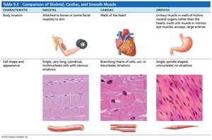
Characteristics and Functions of Muscle Tissue
Excitability: Ability to receive and respond to stimuli.
Contractility: Ability to shorten forcibly when stimulated.
Extensibility: Ability to be stretched.
Elasticity: Ability to recoil to resting length.
Functions include producing movement, maintaining posture, stabilizing joints, and generating heat.
Muscle Cell Physiology
Membrane Potentials and Ion Channels
Muscle contraction is initiated by electrical signals (action potentials) that travel along the muscle cell membrane. These signals are generated by the movement of ions across the membrane through specialized proteins called ion channels.
Resting Membrane Potential (RMP): The voltage difference across the cell membrane at rest, typically -70 to -90 mV in muscle cells.
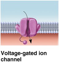
Ion Channels: Integral membrane proteins that allow specific ions (Na+, K+, Ca2+) to move across the membrane. Channels can be leaky (always open) or gated (open in response to stimuli).
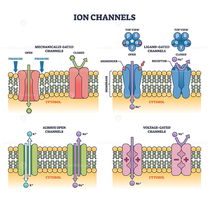
Action Potential Phases
Depolarization: Opening of Na+ channels causes influx of Na+, making the inside of the cell more positive.
Repolarization: Na+ channels close, K+ channels open, and K+ exits the cell, restoring negativity inside.
Hyperpolarization: Sometimes, K+ efflux overshoots, making the cell more negative than RMP before returning to baseline.
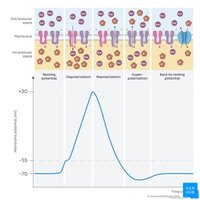
Gated Ion Channels in Muscle Cells
Chemically Gated Channels: Open in response to neurotransmitters (e.g., acetylcholine at the neuromuscular junction).
Voltage-Gated Channels: Open or close in response to changes in membrane potential, propagating the action potential along the muscle fiber.
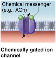
Muscle Structure and Organization
Gross Structure of Skeletal Muscle
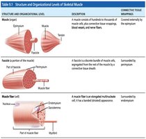
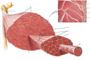
Skeletal muscle is an organ composed of muscle fibers, connective tissue, blood vessels, and nerves. It is organized into bundles for efficient force generation and control.
Epimysium: Surrounds the entire muscle.
Perimysium: Surrounds bundles of muscle fibers called fascicles.
Endomysium: Surrounds individual muscle fibers.
Muscle Attachments
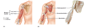
Origin: Attachment to immovable or less movable bone.
Insertion: Attachment to movable bone.
Direct Attachment: Epimysium fused to periosteum or perichondrium.
Indirect Attachment: Connective tissue wrappings extend as tendon or aponeurosis.
Microanatomy of Muscle Fibers
Sarcolemma: Plasma membrane of muscle fiber.
Sarcoplasm: Cytoplasm of muscle fiber, containing glycosomes and myoglobin.
Myofibrils: Rodlike structures that make up most of the muscle cell volume and contain the contractile elements.
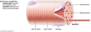
Sarcoplasmic Reticulum and T Tubules
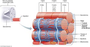
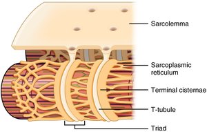
Sarcoplasmic Reticulum (SR): Stores and releases Ca2+ for muscle contraction.
T Tubules: Invaginations of the sarcolemma that transmit action potentials deep into the muscle fiber.
Triad: A T tubule flanked by two terminal cisterns of the SR, crucial for excitation-contraction coupling.
Myofibril Structure and Sarcomere Organization
Myofilaments
Thick Filaments: Composed of myosin molecules with heads that form cross-bridges with actin during contraction.
Thin Filaments: Composed of actin, tropomyosin, and troponin. Actin provides binding sites for myosin heads; tropomyosin blocks these sites at rest; troponin binds Ca2+ to initiate contraction.
Elastic Filaments: Composed of titin, which helps maintain the structure and elasticity of the sarcomere.
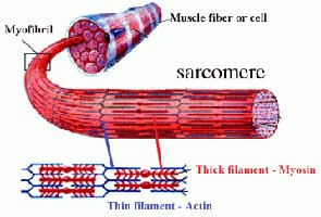
Sarcomere: The Contractile Unit
The sarcomere is the smallest functional unit of muscle fiber, defined as the region between two Z discs. It contains overlapping thick and thin filaments, which give rise to the striated appearance of skeletal and cardiac muscle.
A Band: Dark region containing thick filaments.
I Band: Light region containing thin filaments only.
H Zone: Lighter region in the middle of the A band where there are only thick filaments.
M Line: Line of proteins that holds thick filaments together in the H zone.
Z Disc: Anchors thin filaments and defines the boundaries of a sarcomere.
Sliding Filament Model of Contraction
During contraction, thin filaments slide past thick filaments, shortening the sarcomere without changing the length of the filaments themselves. This process is powered by ATP and regulated by Ca2+ release from the SR.
Muscle Contraction Physiology
Neuromuscular Junction (NMJ) and Muscle Fiber Excitation
NMJ: The synapse between a motor neuron and a muscle fiber, where acetylcholine (ACh) is released to initiate muscle contraction.
Excitation-Contraction Coupling: The process by which an action potential in the sarcolemma leads to Ca2+ release from the SR and subsequent muscle contraction.
Cross Bridge Cycling
Cross bridge formation: Myosin head attaches to actin.
Power stroke: Myosin head pivots, pulling actin toward the M line.
Cross bridge detachment: ATP binds to myosin, causing it to detach from actin.
Cocking of myosin head: ATP hydrolysis re-energizes the myosin head.
This cycle repeats as long as Ca2+ and ATP are available.
Energy for Muscle Contraction
ATP Sources
Direct Phosphorylation: Creatine phosphate donates a phosphate to ADP to form ATP.
Anaerobic Pathway: Glycolysis produces ATP and lactic acid without oxygen.
Aerobic Pathway: Glucose and fatty acids are broken down in the presence of oxygen to produce ATP.
ATP is required for cross bridge cycling, Ca2+ pumping, and restoring ion gradients after contraction.
Muscle Twitch and Graded Responses
Muscle Twitch
A muscle twitch is the response of a muscle fiber to a single action potential. It consists of three phases:
Latent Period: Events of excitation-contraction coupling.
Contraction Period: Cross bridge formation and tension increase.
Relaxation Period: Ca2+ reentry into SR and tension decline.
Graded Muscle Responses
Temporal Summation: Increased frequency of stimulation increases force of contraction.
Recruitment: Increased number of motor units activated increases force.
Muscle Fiber Types and Adaptation
Muscle Fiber Types
Slow Oxidative Fibers: Endurance, slow contraction, fatigue-resistant.
Fast Oxidative Fibers: Intermediate contraction speed and fatigue resistance.
Fast Glycolytic Fibers: Fast contraction, powerful, fatigue quickly.
Adaptation to Exercise
Aerobic Exercise: Increases capillaries, mitochondria, and myoglobin; enhances endurance.
Resistance Exercise: Increases muscle size (hypertrophy) and strength.
Smooth Muscle
Structure and Function
Found in walls of hollow organs (except heart).
Spindle-shaped, single nucleus, non-striated.
Arranged in longitudinal and circular layers for coordinated contraction.
Differences from Skeletal Muscle
No sarcomeres, T tubules, or troponin; uses calmodulin for Ca2+ binding.
Gap junctions allow coordinated contraction.
Contraction is slower, more sustained, and energy-efficient.
Regulation of Contraction
Controlled by autonomic nerves, hormones, and local factors.
Can contract in response to stretch, chemical signals, or spontaneously (pacemaker cells).
Special Features
Stress-relaxation response allows organs to stretch and adapt to new volumes.
Can contract when stretched to half or twice its resting length.
