 Back
BackMuscles and Muscle Tissue: Structure, Function, and Contraction
Study Guide - Smart Notes
 Tailored notes based on your materials, expanded with key definitions, examples, and context.
Muscle Tissue: Overview and Types
Muscle Tissue Terminology and Characteristics
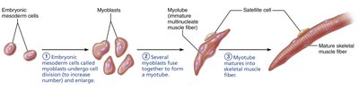
Muscle tissue is essential for movement, posture, joint stability, and heat generation. The prefixes myo-, mys-, and sarco- refer to muscle. Sarcoplasm is the cytoplasm of a muscle cell, and myocardium refers to heart muscle. Muscle fibers are elongated cells found in skeletal and smooth muscle, but not in cardiac muscle, which is branched. Muscle fibers are multinucleate due to the fusion of myoblasts during development.
Excitability: Ability to receive and respond to stimuli.
Contractility: Ability to shorten forcibly when stimulated.
Extensibility: Ability to be stretched and relaxed.
Elasticity: Ability to recoil to resting length.
Muscle tissue functions include producing movement, maintaining posture, stabilizing joints, and generating heat.
Types of Muscle Tissue
There are three types of muscle tissue: skeletal, cardiac, and smooth. Each type has distinct structural and functional characteristics.
Characteristic | Skeletal | Cardiac | Smooth |
|---|---|---|---|
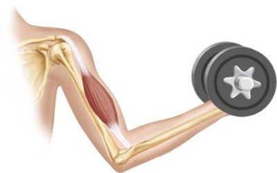
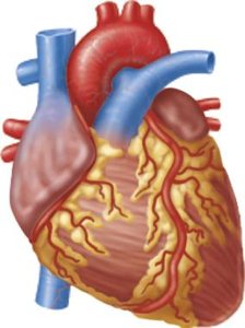
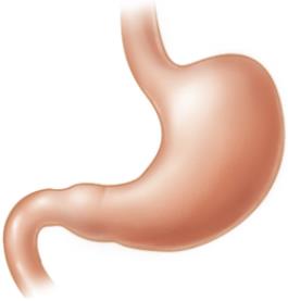
Body Location | Attached to bones or skin | Walls of the heart | Walls of hollow organs (except heart) |
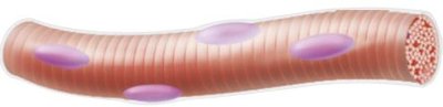
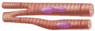
Cell Shape & Appearance | Long, cylindrical, multinucleate, striated | Branching chains, uni- or binucleate, striated | Spindle-shaped, uninucleate, no striations |
Structure of Skeletal Muscle
Connective Tissue Sheaths
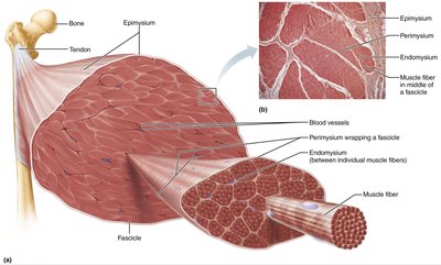
Skeletal muscle is organized into bundles and surrounded by connective tissue sheaths:
Epimysium: Dense irregular connective tissue, surrounds the entire muscle.
Perimysium: Dense irregular connective tissue, surrounds fascicles (bundles of muscle fibers).
Endomysium: Areolar connective tissue, surrounds individual muscle fibers.
Muscle Attachments
Muscles attach to bones at two points:
Origin: Attachment to immovable or less movable bone.
Insertion: Attachment to movable bone.
Attachments can be direct (muscle to bone) or indirect (via tendons or aponeuroses).
Skeletal Muscle Fiber Structure
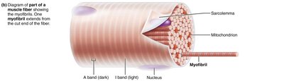
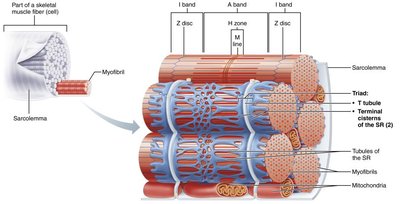
Skeletal muscle fibers are long, cylindrical, multinucleate cells. The sarcolemma is the plasma membrane, and the sarcoplasm is the cytoplasm, containing glycosomes (glycogen storage) and myoglobin (oxygen storage). Specialized structures include myofibrils, sarcoplasmic reticulum, and T tubules.
Myofibrils and Sarcomeres
Myofibril Structure
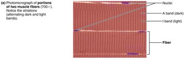
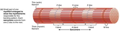
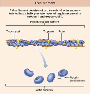
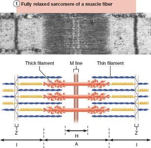
Myofibrils are rod-like elements that make up most of the muscle cell volume. They contain repeating units called sarcomeres, the smallest contractile units of muscle. Striations are due to the arrangement of thick (myosin) and thin (actin) filaments.
Sarcomere Organization
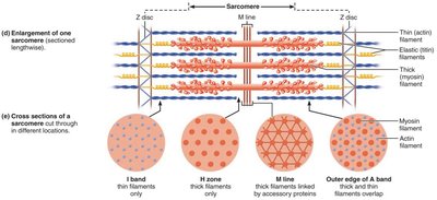
A sarcomere extends from one Z disc to the next. Thick filaments are anchored to the M line and indirectly to Z discs by titin protein.
Myofilaments: Actin and Myosin
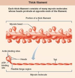
Myofilaments are organized within the sarcomere:
Actin: Thin filaments
Myosin: Thick filaments
Sarcoplasmic Reticulum and T Tubules
The sarcoplasmic reticulum (SR) is a network of smooth endoplasmic reticulum surrounding each myofibril, storing calcium ions. T tubules are invaginations of the sarcolemma that conduct impulses deep into the muscle fiber. The triad consists of a T tubule and two terminal cisterns of the SR.
Sliding Filament Model of Contraction
Mechanism of Contraction
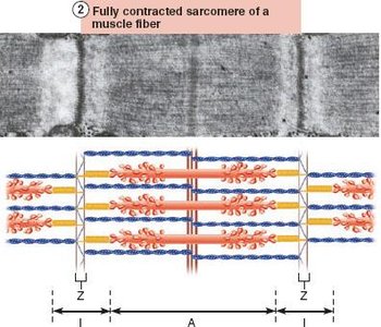
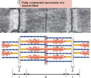
During contraction, thin filaments slide past thick filaments, increasing their overlap but not changing their length. This sliding shortens the muscle fiber.
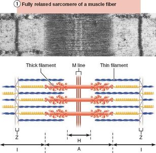
I bands shorten
Z discs move closer together
H zones disappear
A bands move closer together
Excitation-Contraction Coupling
Ion Channels in Muscle Contraction
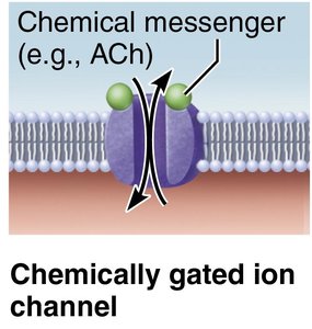
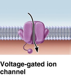
Muscle contraction is regulated by ion channels:
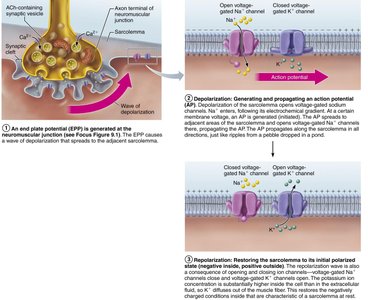
Chemically gated ion channels: Open in response to neurotransmitters (e.g., acetylcholine).
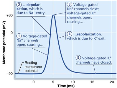
Voltage-gated ion channels: Open in response to changes in membrane potential.
Neuromuscular Junction (NMJ)
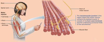
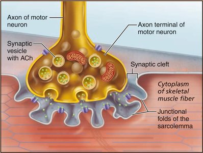
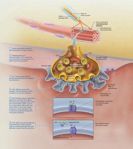
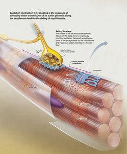
The NMJ is where the motor neuron meets the skeletal muscle fiber. The axon terminal releases acetylcholine (ACh) into the synaptic cleft, which binds to receptors on the sarcolemma, initiating muscle contraction.
Steps of Skeletal Muscle Contraction
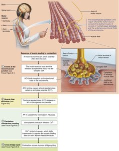
Events at the neuromuscular junction: Action potential arrives, Ca2+ channels open, ACh is released, binds to receptors, and triggers end plate potential.
Muscle fiber excitation: End plate potential triggers an action potential across the sarcolemma.
Excitation-contraction coupling: Action potential travels down T tubules, causing Ca2+ release from SR, which binds to troponin, exposing myosin binding sites.
Cross bridge cycle: Myosin heads bind to actin, perform power stroke, detach with ATP, and reset.
Muscle Twitch and Graded Contractions
Muscle Twitch
A muscle twitch is the response of a muscle fiber to a single action potential. It consists of three phases:
Latent period: Excitation-contraction coupling
Contraction period: Cross bridge formation
Relaxation period: Ca2+ reentry into SR
Graded Muscle Contractions
Muscle contractions are graded by changing the frequency and strength of stimulation. Temporal (wave) summation occurs when stimuli are delivered in rapid succession, leading to increased force. Sustained contractions can result in unfused (incomplete) or fused (complete) tetanus.
Muscle Fiber Types
Types of Skeletal Muscle Fibers
Characteristic | Slow Oxidative | Fast Oxidative | Fast Glycolytic |
|---|---|---|---|
Speed of contraction | Slow | Fast | Fast |
ATP synthesis | Aerobic | Aerobic/anaerobic | Anaerobic glycolysis |
Myoglobin | High | High | Low |
Fatigue resistance | High | Intermediate | Low |
Best for | Endurance | Sprinting/walking | Short, powerful movements |
Smooth Muscle
Structure and Function
Smooth muscle is found in the walls of hollow organs. It has two layers: longitudinal (shortens organ) and circular (constricts lumen). Smooth muscle cells are spindle-shaped, uninucleate, and lack striations.
Comparison of Skeletal and Smooth Muscle
Characteristic | Skeletal | Smooth |
|---|---|---|
Connective tissue | Epimysium, perimysium, endomysium | Endomysium only |
Myofibrils/sarcomeres | Yes | No (dense bodies instead) |
T tubules | Yes | No (caveolae instead) |
SR | Elaborate | Less developed |
Gap junctions | No | Yes (unitary muscle) |
Regulation | Voluntary (somatic NS) | Involuntary (autonomic NS, hormones, stretch) |
Calcium source | SR | Extracellular fluid and SR |
Calcium regulation | Troponin | Calmodulin |
Contraction speed | Fast | Very slow |
