 Back
BackMuscles and Muscle Tissue: Structure, Function, and Physiology
Study Guide - Smart Notes
 Tailored notes based on your materials, expanded with key definitions, examples, and context.
Muscle Tissue Overview
Introduction to Muscle Tissue
Muscle tissue is essential for movement, posture, and various physiological processes. It comprises nearly half of the body's mass and is specialized for converting chemical energy (ATP) into mechanical energy, enabling force generation and movement.
Key prefixes: myo-, mys-, and sarco- refer to muscle structures (e.g., sarcoplasm = muscle cell cytoplasm).
Three types of muscle tissue: Skeletal, Cardiac, Smooth.
Types of Muscle Tissue
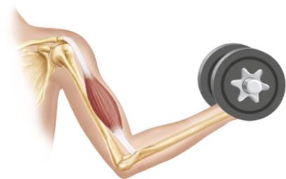
Skeletal Muscle: Attached to bones and skin, voluntary, striated, contracts rapidly but tires easily. Responsible for body movement and posture.
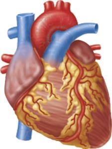
Cardiac Muscle: Found only in the heart, involuntary, striated, contracts at a steady rate set by pacemaker cells but modulated by the nervous system.
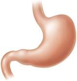
Smooth Muscle: Found in walls of hollow organs (e.g., stomach, bladder, airways), involuntary, non-striated, responsible for moving substances through internal body channels.
Characteristic | Skeletal | Cardiac | Smooth |
|---|---|---|---|
Location | Attached to bones/skin | Heart walls | Walls of hollow organs |
Control | Voluntary | Involuntary | Involuntary |
Striations | Yes | Yes | No |
Cell Shape | Long, cylindrical, multinucleate | Branching, uni/binucleate | Spindle-shaped, uninucleate |
Characteristics of Muscle Tissue
Excitability: Ability to receive and respond to stimuli.
Contractility: Ability to shorten forcibly when stimulated.
Extensibility: Ability to be stretched.
Elasticity: Ability to recoil to resting length.
Functions of Muscle Tissue
Produce movement (locomotion, manipulation, pumping blood, digestion)
Maintain posture and body position
Stabilize joints
Generate heat during contraction
Skeletal Muscle Structure and Organization
Components of Skeletal Muscle
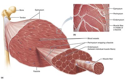
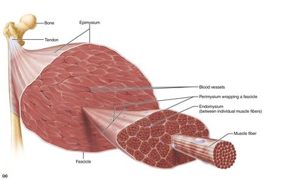
Skeletal muscle is an organ composed of muscle fibers, connective tissue, blood vessels, and nerves. It is organized into several hierarchical levels:
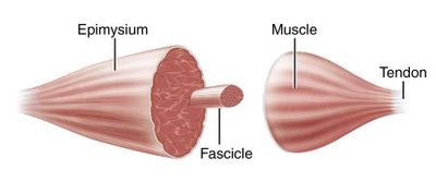
Muscle (organ): Surrounded by epimysium.
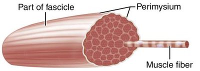
Fascicle: Bundle of muscle fibers, surrounded by perimysium.
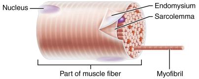
Muscle fiber (cell): Surrounded by endomysium.
Level | Description | Connective Tissue |
|---|---|---|
Muscle | Hundreds to thousands of muscle cells, blood vessels, nerves | Epimysium |
Fascicle | Bundle of muscle fibers | Perimysium |
Muscle fiber | Elongated, multinucleate cell | Endomysium |
Attachments
Direct (fleshy): Epimysium fused to periosteum of bone or perichondrium of cartilage.
Indirect: Connective tissue wrappings extend beyond muscle as a tendon or aponeurosis.
Microscopic Anatomy of Skeletal Muscle Fibers
Muscle Fiber Structure
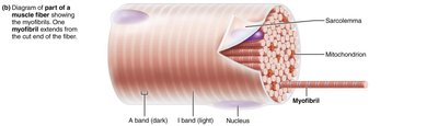
Sarcolemma: Plasma membrane of muscle fiber.
Sarcoplasm: Cytoplasm containing glycosomes (glycogen storage) and myoglobin (O2 storage).
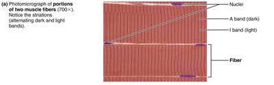
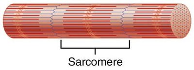
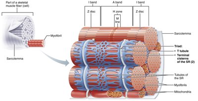
Myofibrils: Densely packed, rodlike elements responsible for muscle contraction and striations.
Sarcomere and Myofilaments
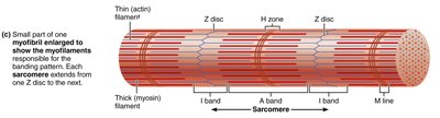
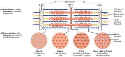
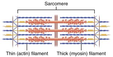
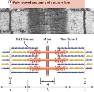
Sarcomere: Smallest contractile unit, extends from Z disc to Z disc.
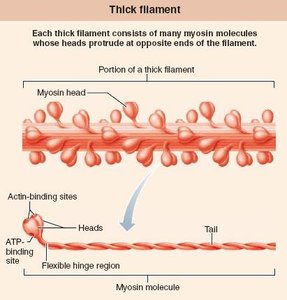
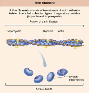
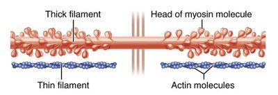
Myofilaments: Thick (myosin) and thin (actin) filaments arranged in a hexagonal pattern.
Striations: Alternating A bands (dark) and I bands (light) due to arrangement of myofilaments.
Specialized Proteins
Myosin: Thick filament with heads that form cross bridges during contraction.
Actin: Thin filament with binding sites for myosin heads; includes regulatory proteins tropomyosin and troponin.
Titin: Elastic filament that maintains sarcomere structure and elasticity.
Dystrophin: Links thin filaments to sarcolemma proteins; mutations cause Duchenne muscular dystrophy.
Excitation-Contraction Coupling and Muscle Contraction
Sarcoplasmic Reticulum and T Tubules
Sarcoplasmic Reticulum (SR): Stores and releases Ca2+ for muscle contraction.
T Tubules: Invaginations of sarcolemma that transmit action potentials deep into muscle fiber.
Triad: Structure formed by a T tubule and two terminal cisterns of the SR.
Sliding Filament Model of Contraction
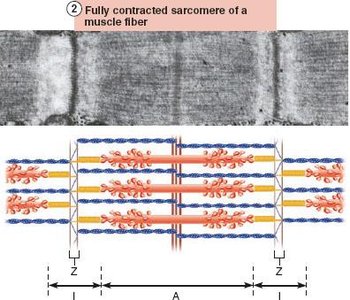
Muscle contraction occurs when myosin heads bind to actin, forming cross bridges and pulling thin filaments toward the center of the sarcomere. This process shortens the muscle fiber without changing the length of the filaments.
During contraction: I bands shorten, Z discs move closer, H zones disappear, A bands move closer together.
Neuromuscular Junction and Action Potential
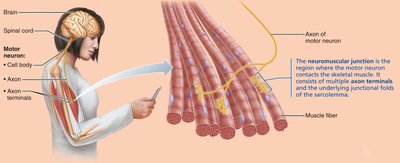
Motor Neurons: Stimulate muscle fibers via the neuromuscular junction (NMJ).
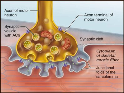
Neurotransmitter: Acetylcholine (ACh) is released from the axon terminal, binds to receptors on the sarcolemma, and initiates an action potential.
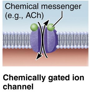
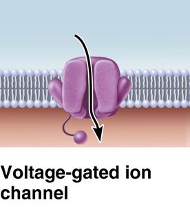
Ion Channels: Chemically gated (ACh) and voltage-gated channels regulate ion flow and membrane potential changes.
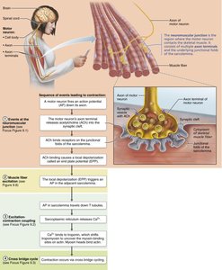
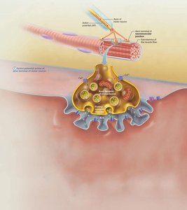
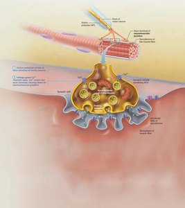
Steps in Muscle Fiber Excitation and Contraction
Action potential arrives at axon terminal.
Voltage-gated Ca2+ channels open; Ca2+ enters neuron.
ACh is released into synaptic cleft.
ACh binds to receptors, opening Na+ channels and generating end plate potential.
Action potential propagates along sarcolemma and down T tubules.
Ca2+ is released from SR, initiating contraction.
Cross Bridge Cycle
Requires Ca2+ and ATP.
Four steps: Cross bridge formation, power stroke, cross bridge detachment, cocking of myosin head.
Whole Muscle Contraction
Motor Units
A motor unit consists of a motor neuron and all the muscle fibers it innervates.
Smaller motor units allow for fine control; larger units generate more force.
Muscle Twitch and Graded Responses
Muscle Twitch: Response to a single stimulus; consists of latent, contraction, and relaxation phases.
Graded Responses: Varying strength of contraction by changing stimulus frequency (temporal summation) or strength (recruitment).
Types of Contractions
Isotonic: Muscle changes length (concentric = shortens, eccentric = lengthens).
Isometric: Muscle tension increases but does not change length.
Energy for Muscle Contraction
ATP Regeneration Pathways
Direct phosphorylation: Creatine phosphate donates phosphate to ADP to form ATP.
Anaerobic pathway: Glycolysis and lactic acid formation (no oxygen required).
Aerobic pathway: Glycolysis followed by aerobic respiration in mitochondria (requires oxygen).
Equation for direct phosphorylation:
Muscle Fatigue and Recovery
Fatigue results from ionic imbalances, increased inorganic phosphate, decreased ATP, and other factors.
Recovery involves replenishing oxygen, ATP, and glycogen stores (EPOC: excess postexercise oxygen consumption).
Muscle Fiber Types and Adaptations
Muscle Fiber Types
Slow oxidative fibers: Endurance, fatigue-resistant, aerobic metabolism.
Fast oxidative fibers: Intermediate properties, aerobic and some anaerobic capacity.
Fast glycolytic fibers: Short-term, powerful movements, fatigue quickly, anaerobic metabolism.
Type | Contraction Speed | ATP Pathway | Fatigue Resistance | Best Suited For |
|---|---|---|---|---|
Slow Oxidative | Slow | Aerobic | High | Endurance, posture |
Fast Oxidative | Fast | Aerobic/Some Anaerobic | Intermediate | Sprinting, walking |
Fast Glycolytic | Fast | Anaerobic | Low | Short, intense movements |
Muscle Adaptation to Exercise
Aerobic exercise: Increases capillaries, mitochondria, myoglobin; enhances endurance.
Resistance exercise: Increases muscle size (hypertrophy), strength, and connective tissue.
Disuse atrophy: Muscle wasting due to inactivity or loss of neural stimulation.
Smooth Muscle
Structure and Function
Found in walls of hollow organs (except heart).
Spindle-shaped, uninucleate, non-striated cells; organized in sheets.
Contraction is involuntary, slow, and can be sustained for long periods.
Differences from Skeletal Muscle
No sarcomeres, T tubules, or troponin; uses calmodulin for Ca2+ binding.
Gap junctions allow for coordinated contraction.
Contraction mechanism involves Ca2+ entry from extracellular space, activation of calmodulin, and phosphorylation of myosin.
Types of Smooth Muscle
Unitary (visceral): Most common, found in hollow organs, electrically coupled by gap junctions.
Multi-unit: Found in large airways, arteries, arrector pili, and iris; fibers act independently.
Development and Aging of Muscle Tissue
All muscle types develop from embryonic mesoderm (myoblasts).
Skeletal muscle fibers form by fusion of myoblasts; cardiac and smooth muscle develop gap junctions.
Muscle mass and function decline with age (sarcopenia), but regular exercise can slow this process.
