 Back
BackMuscles and Muscle Tissue: Structure, Function, and Physiology
Study Guide - Smart Notes
 Tailored notes based on your materials, expanded with key definitions, examples, and context.
Muscle Tissue Overview
Types of Muscle Tissue
Muscle tissue is essential for movement and comprises nearly half of the body's mass. It transforms chemical energy (ATP) into mechanical energy, enabling force generation. There are three primary types of muscle tissue, each with distinct structural and functional characteristics:
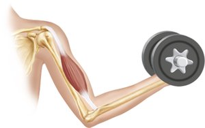
Skeletal Muscle: Attached to bones and skin, responsible for voluntary movements. Fibers are long, multinucleated, and striated.
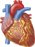
Cardiac Muscle: Found only in the heart, responsible for pumping blood. Fibers are striated, involuntary, and have a single nucleus.
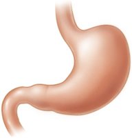
Smooth Muscle: Located in walls of hollow organs (e.g., stomach, bladder, airways). Fibers are non-striated, involuntary, and have a single nucleus.
Muscle Type | Location | Control | Striations | Nucleus |
|---|---|---|---|---|
Skeletal | Bones, skin | Voluntary | Yes | Multiple |
Cardiac | Heart | Involuntary | Yes | Single |
Smooth | Hollow organs | Involuntary | No | Single |
Characteristics of Muscle Tissue
All muscle tissues share four main properties:
Excitability: Ability to respond to stimuli.
Contractility: Ability to shorten forcibly.
Extensibility: Ability to be stretched.
Elasticity: Ability to return to resting length.
Functions of Muscle Tissue
Produce movement: Locomotion and manipulation.
Maintain posture: Stabilizes body position.
Stabilize joints: Reinforces joint stability.
Generate heat: Maintains body temperature.
Skeletal Muscle Structure
Connective Tissue Sheaths
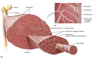
Skeletal muscle is organized into layers of connective tissue that support and reinforce the muscle:
Epimysium: Surrounds entire muscle.
Perimysium: Surrounds fascicles (bundles of fibers).
Endomysium: Surrounds individual muscle fibers.
Skeletal Muscle Fiber Anatomy
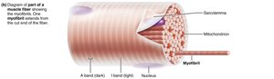
Skeletal muscle fibers are long, cylindrical cells with multiple nuclei. Key structures include:
Sarcolemma: Plasma membrane of muscle fiber.
Sarcoplasm: Cytoplasm containing glycosomes (glycogen storage) and myoglobin (oxygen storage).
Myofibrils: Rodlike elements responsible for striations and contraction.
Sarcoplasmic Reticulum (SR): Stores and releases calcium ions.
T Tubules: Invaginations of sarcolemma that transmit electrical signals.
Myofibril Structure and Function
Striations and Sarcomeres
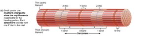
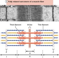
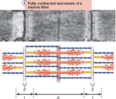
Striations are formed by alternating dark (A bands) and light (I bands) regions. The sarcomere is the functional unit of muscle contraction, defined as the segment between two Z discs.
A Band: Contains thick filaments (myosin).
I Band: Contains thin filaments (actin).
H Zone: Lighter region in the middle of A band.
M Line: Protein line bisecting H zone.
Z Disc: Anchors thin filaments.
Myofilaments
Myofilaments are organized within the sarcomere:
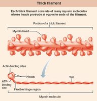
Thick Filaments: Composed of myosin molecules with heads that form cross bridges during contraction.
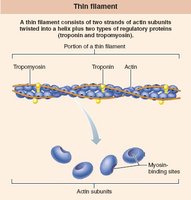
Thin Filaments: Composed of actin, tropomyosin, and troponin. Actin provides binding sites for myosin heads.
Elastic Filament: Made of titin, maintains sarcomere structure.
Muscle Contraction Mechanisms
Sliding Filament Model
Muscle contraction occurs when thin filaments slide past thick filaments, increasing overlap. The process is initiated by cross bridge formation between myosin heads and actin.
Neither filament changes length; overlap increases.
Contraction ends when cross bridges become inactive.
Excitation-Contraction Coupling
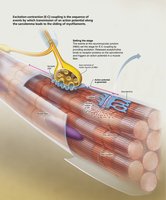
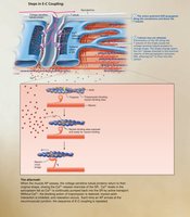
Excitation-contraction coupling links the electrical signal (action potential) to muscle contraction:
Action potential travels along sarcolemma and T tubules.
Triggers release of Ca2+ from SR.
Ca2+ binds to troponin, moving tropomyosin and exposing actin binding sites.
Myosin heads bind to actin, initiating contraction.
Cross Bridge Cycle
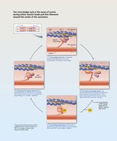
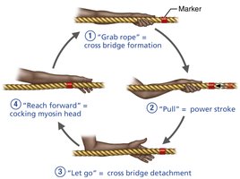
The cross bridge cycle consists of four steps:
Cross bridge formation: Myosin head attaches to actin.
Power stroke: Myosin head pivots, pulling actin toward M line.
Detachment: ATP binds to myosin, causing detachment from actin.
Cocking: ATP hydrolysis re-energizes myosin head.
Motor Units and Muscle Twitch
Motor Unit
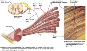
A motor unit consists of a motor neuron and all the muscle fibers it innervates. Smaller motor units allow finer control.
Muscle Twitch
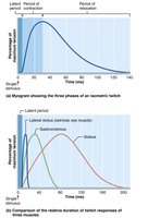
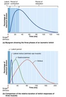
A muscle twitch is the response of a muscle fiber to a single action potential. It consists of three phases:
Latent period: Excitation-contraction coupling.
Contraction period: Cross bridge formation and tension increase.
Relaxation period: Ca2+ reentry into SR and tension decline.
Graded Muscle Contractions
Temporal Summation and Tetanus
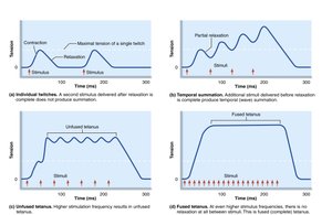
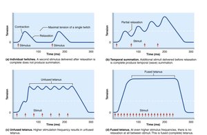
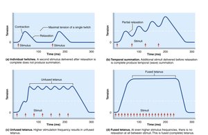
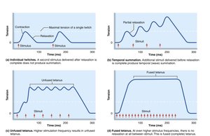
Graded muscle contractions allow variation in strength and smoothness. Temporal summation occurs when stimuli are delivered rapidly, leading to increased force. If frequency is high enough, contractions fuse into tetanus.
Recruitment and Size Principle
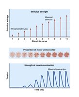
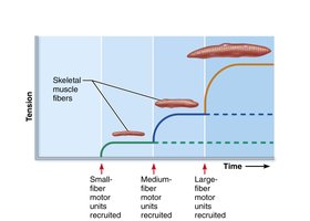
Recruitment involves activating more motor units to increase contraction strength. The size principle states that smaller motor units are recruited first, followed by larger ones as intensity increases.
Muscle Tone and Types of Contractions
Muscle Tone
Muscle tone is a constant, slightly contracted state maintained by spinal reflexes, keeping muscles ready for action.
Isotonic and Isometric Contractions
Muscle contractions can be:
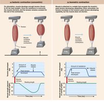
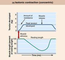
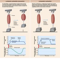
Isotonic: Muscle changes length and moves load (concentric: shortens; eccentric: lengthens).
Isometric: Muscle tension increases but does not change length.
ATP and Muscle Contraction
ATP Regeneration Pathways
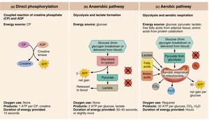
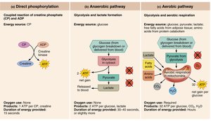
ATP is essential for muscle contraction and is regenerated by three mechanisms:
Direct phosphorylation: Creatine phosphate donates phosphate to ADP.
Anaerobic pathway: Glycolysis and lactate formation (no oxygen required).
Aerobic pathway: Glycolysis and aerobic respiration in mitochondria (oxygen required).
Energy Use During Exercise
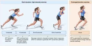
Short-duration, high-intensity exercise relies on stored ATP and creatine phosphate, while prolonged exercise uses aerobic pathways.
Factors Affecting Muscle Contraction
Force of Contraction
The force generated depends on:
Frequency of stimulation
Number of fibers recruited
Size of muscle fibers
Degree of muscle stretch (length-tension relationship)
Velocity and Duration
Contraction speed and duration are influenced by:
Muscle fiber type (slow oxidative, fast oxidative, fast glycolytic)
Load
Recruitment
Muscle Response to Exercise
Aerobic and Resistance Exercise
Aerobic exercise increases endurance, capillaries, mitochondria, and myoglobin. Resistance exercise leads to hypertrophy and increased strength.
Smooth Muscle Structure and Function
Organization and Differences
Smooth muscle is found in hollow organs and organized into sheets. It differs from skeletal muscle in cell shape, innervation, and contraction mechanism.
Spindle-shaped, single nucleus, no striations
Innervated by autonomic nervous system
Uses calmodulin instead of troponin for Ca2+ binding
Types of Smooth Muscle
Unitary (visceral): Found in most hollow organs, electrically coupled by gap junctions.
Multi unit: Found in large airways, arteries, arrector pili, and iris; independent fibers, graded contractions.
Developmental Aspects
Muscle Development and Regeneration
All muscle tissues develop from myoblasts. Skeletal muscle cells form by fusion, while cardiac and smooth muscle cells develop gap junctions. Regeneration varies among muscle types.
Additional info: This summary expands on brief points with academic context, definitions, and examples for clarity and completeness.
