 Back
BackMuscles and Muscle Tissue: Structure, Function, and Physiology
Study Guide - Smart Notes
 Tailored notes based on your materials, expanded with key definitions, examples, and context.
Muscle Tissue Overview
Introduction to Muscle Tissue
Muscle tissue constitutes nearly half of the body's mass and is essential for transforming chemical energy (ATP) into mechanical energy, enabling force generation and movement. The prefixes myo-, mys-, and sarco- are commonly used in muscle terminology, such as sarcoplasm (muscle cell cytoplasm).
Types of Muscle Tissue
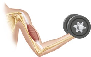
Skeletal Muscle: Attached to bones and skin, skeletal muscle fibers are the longest and have striations. They are voluntary and contract rapidly but tire easily.
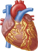
Cardiac Muscle: Found only in the heart, cardiac muscle is striated and involuntary. It contracts at a steady rate due to the heart's pacemaker.
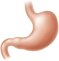
Smooth Muscle: Located in the walls of hollow organs (e.g., stomach, bladder, airways), smooth muscle is non-striated and involuntary.
Muscle Type | Location | Striations | Control |
|---|---|---|---|
Skeletal | Bones, skin | Yes | Voluntary |
Cardiac | Heart | Yes | Involuntary |
Smooth | Hollow organs | No | Involuntary |
Characteristics of Muscle Tissue
Excitability: Ability to respond to stimuli.
Contractility: Ability to shorten forcibly.
Extensibility: Ability to be stretched.
Elasticity: Ability to recoil to resting length.
Functions of Muscle Tissue
Produce movement (locomotion, manipulation)
Maintain posture and body position
Stabilize joints
Generate heat during contraction
Skeletal Muscle Structure
Organizational Structure
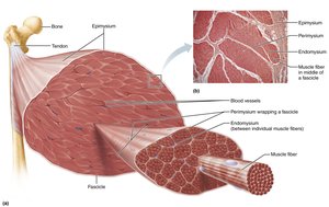
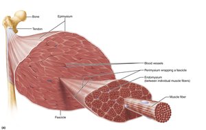
Skeletal muscle is an organ composed of muscle fibers, nerves, blood vessels, and connective tissue sheaths. Each muscle receives a nerve, artery, and veins, ensuring adequate oxygen and nutrient supply and waste removal.
Connective Tissue Sheaths
Epimysium: Surrounds entire muscle; dense irregular connective tissue.
Perimysium: Surrounds fascicles (groups of muscle fibers); fibrous connective tissue.
Endomysium: Surrounds each muscle fiber; fine areolar connective tissue.
Muscle Attachments
Direct (fleshy) attachment: Epimysium fused to periosteum or perichondrium.
Indirect attachment: Connective tissue extends beyond muscle as tendon or aponeurosis.
Skeletal Muscle Fiber Anatomy
Microscopic Anatomy
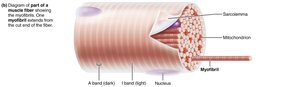
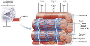
Skeletal muscle fibers are long, cylindrical, multinucleated cells. The sarcolemma is the plasma membrane, and the sarcoplasm is the cytoplasm, rich in glycosomes and myoglobin.
Myofibrils: Densely packed rodlike elements responsible for striations.
Sarcoplasmic Reticulum: Smooth ER network regulating calcium storage and release.
T Tubules: Extensions of sarcolemma that transmit electrical impulses.
Striations and Sarcomeres
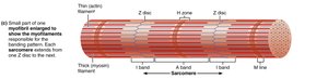
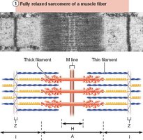
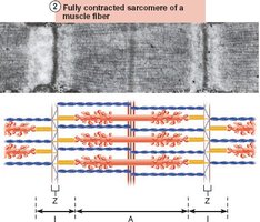
Striations are formed by alternating dark (A bands) and light (I bands) regions. The sarcomere is the smallest contractile unit, defined by the area between two Z discs.
Myofilaments
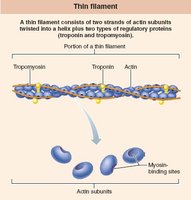
Actin (thin filaments): Anchored to Z discs, composed of G actin and F actin, with regulatory proteins tropomyosin and troponin.
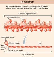
Myosin (thick filaments): Composed of myosin molecules with heads forming cross bridges during contraction.
Other Structural Proteins
Titin: Provides elasticity and holds thick filaments in place.
Dystrophin: Links thin filaments to sarcolemma proteins.
Nebulin, myomesin, C proteins: Maintain sarcomere alignment.
Muscle Contraction Mechanisms
Sarcoplasmic Reticulum and T Tubules
The sarcoplasmic reticulum (SR) stores and releases calcium, while T tubules transmit action potentials deep into the muscle fiber. The triad consists of a T tubule flanked by two terminal cisterns of the SR.
Sliding Filament Model
During contraction, thin filaments slide past thick filaments, increasing overlap. Cross bridges form and break, pulling thin filaments toward the center of the sarcomere, resulting in muscle shortening.
Neuromuscular Junction and Muscle Excitation
Neuromuscular Junction
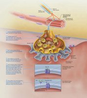
The neuromuscular junction is where the motor neuron communicates with the muscle fiber. Acetylcholine (ACh) is released from the neuron, binds to receptors on the sarcolemma, and initiates an action potential.
Action Potential Generation
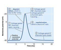
Depolarization: Sodium influx makes the inside of the cell more positive.
Repolarization: Potassium efflux restores resting membrane potential.
Refractory Period: The cell cannot be stimulated again until repolarization is complete.
Excitation-Contraction Coupling
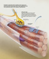
Excitation-contraction coupling links the action potential to muscle contraction. The action potential travels along the sarcolemma and T tubules, triggering calcium release from the SR, which initiates contraction.
Cross Bridge Cycle
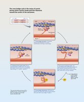
Cross bridge formation: Myosin head binds to actin.
Power stroke: Myosin head pivots, pulling actin.
Detachment: ATP binds to myosin, causing release.
Cocking: ATP hydrolysis re-energizes myosin head.
Whole Muscle Contraction
Motor Units
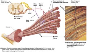
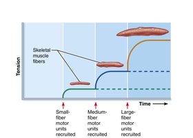
A motor unit consists of a motor neuron and all the muscle fibers it innervates. Smaller motor units allow finer control, and fibers are distributed throughout the muscle.
Muscle Twitch
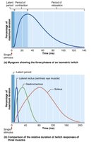
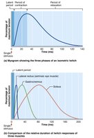
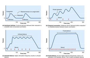
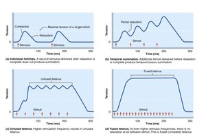
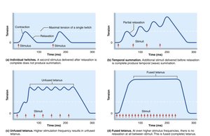
A muscle twitch is the response to a single action potential. It consists of three phases: latent period, contraction, and relaxation.
Graded Muscle Contractions
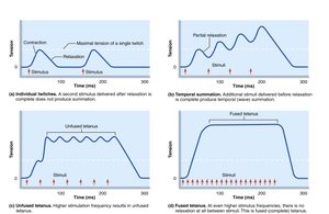
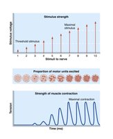
Muscle contractions are graded by changing the frequency and strength of stimulation. Temporal summation and recruitment allow for smooth, controlled movements.
Muscle Tone
Muscle tone is a constant, slightly contracted state maintained by spinal reflexes, keeping muscles ready for action.
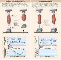
Isotonic and Isometric Contractions
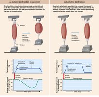
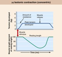
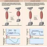
Isotonic: Muscle changes length and moves load (concentric: shortens; eccentric: lengthens).
Isometric: Muscle tension increases but does not exceed load; muscle does not change length.
Energy for Muscle Contraction
ATP Regeneration
ATP is the sole energy source for muscle contraction and must be regenerated rapidly. Three mechanisms are used:
Direct phosphorylation: Creatine phosphate donates phosphate to ADP.
Anaerobic pathway: Glycolysis and lactate formation.
Aerobic pathway: Glycolysis and aerobic respiration in mitochondria.
Muscle Fatigue and Recovery
Muscle fatigue is the inability to contract despite stimulation, caused by ionic imbalances, increased inorganic phosphate, decreased ATP, and glycogen depletion. Recovery requires replenishing oxygen, lactate conversion, glycogen restoration, and ATP/creatine phosphate resynthesis (EPOC).
Factors Affecting Muscle Contraction
Force of Contraction
Frequency of stimulation
Number of fibers stimulated (recruitment)
Size of muscle fibers
Degree of muscle stretch (length-tension relationship)
Velocity and Duration of Contraction
Muscle fiber type: Slow oxidative, fast oxidative, fast glycolytic
Load: Greater load decreases contraction speed and duration
Recruitment: More motor units increase speed and duration
Muscle Response to Exercise and Development
Exercise Effects
Aerobic exercise: Increases capillaries, mitochondria, myoglobin, endurance, and strength.
Resistance exercise: Causes hypertrophy, increased strength, and fiber size.
Muscle Atrophy and Aging
Disuse atrophy: Loss of muscle mass due to inactivity or neural loss.
Sarcopenia: Age-related muscle loss, reversible with exercise.
Developmental Aspects
Muscle tissues develop from myoblasts.
Regeneration is limited in skeletal muscle; smooth muscle regenerates throughout life.
Muscular development in infants reflects neuromuscular coordination.
Additional info: The notes expand on brief points with academic context, definitions, and examples, and include relevant images to reinforce key concepts.
