 Back
BackMuscles and Muscle Tissue: Structure, Function, and Physiology
Study Guide - Smart Notes
 Tailored notes based on your materials, expanded with key definitions, examples, and context.
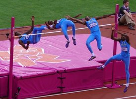
Muscle Functions
Overview of Muscle Functions
Muscles are specialized tissues responsible for producing movement, maintaining posture, generating heat, and stabilizing joints. These functions are essential for the proper functioning of the human body and are achieved through the contraction and relaxation of muscle fibers.
Movement: Muscles move the skeleton, blood, and food through coordinated contractions.
Posture: Muscles maintain posture through continuous, low-level contractions known as muscle tone.
Heat Generation: Muscle contractions produce heat, which helps maintain body temperature.
Joint Stabilization: Muscles stabilize joints, especially during movement, by maintaining tension across the joint.


Muscle Attachment
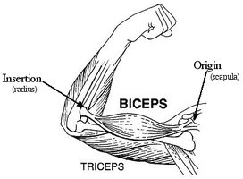
Origin and Insertion
Muscles attach to bones or cartilage at two main points: the origin (fixed attachment) and the insertion (movable attachment). Attachments can be direct or indirect:
Indirect Attachment: Most common; muscle tapers into a tendon that attaches to bone or cartilage.
Direct Attachment: Muscle fibers attach directly to the periosteum or perichondrium without a tendon.
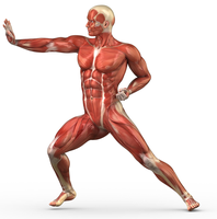
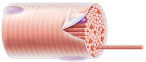
Anatomy of a Muscle
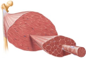
Muscle Structure and Connective Tissue Layers
Muscles are organized into hierarchical structures, each surrounded by connective tissue:
Epimysium: Dense irregular connective tissue surrounding the entire muscle.
Perimysium: Dense irregular connective tissue surrounding each fascicle (bundle of muscle fibers).
Endomysium: Areolar connective tissue surrounding each individual muscle fiber (cell).


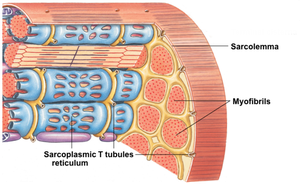
Anatomy of a Muscle Fiber
Muscle Fiber Structure
A muscle fiber is a single, elongated cell containing multiple nuclei and specialized structures for contraction:
Sarcolemma: The excitable cell membrane that conducts electrical signals.
Myofibrils: Rod-like structures within the muscle fiber, composed of repeating units called sarcomeres.
Sarcoplasmic Reticulum (SR): Specialized endoplasmic reticulum that stores and releases Ca2+ ions.
T Tubules: Invaginations of the sarcolemma that transmit electrical signals deep into the muscle fiber.


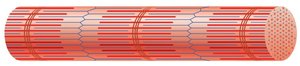
Sarcomere Structure and Function
Organization of Myofilaments
The sarcomere is the functional unit of muscle contraction, composed of overlapping thick (myosin) and thin (actin) filaments. The arrangement of these filaments gives skeletal muscle its striated appearance.
Z disc: Anchors actin filaments; marks the boundary of each sarcomere.
M line: Anchors myosin filaments in the center of the sarcomere.
A band: Dark region where actin and myosin overlap.
I band: Light region containing only actin filaments.
H band: Lighter region within the A band where only myosin is present.


Microstructure of Myofilaments
Thick and Thin Filaments
Myofilaments are protein structures responsible for muscle contraction:
Myosin (thick filament): Composed of ~300 myosin molecules, each with a tail and two heads. Heads have actin binding sites and ATPase activity.
Actin (thin filament): Contains myosin binding sites, covered by tropomyosin. Troponin regulates the position of tropomyosin.
When Ca2+ binds to troponin, tropomyosin moves, exposing the myosin binding sites on actin, allowing cross-bridge formation.
The Cross Bridge Cycle
Mechanism of Muscle Contraction
The cross bridge cycle describes the process by which myosin heads bind to actin, pull the filaments, and generate muscle contraction. The cycle is powered by ATP and regulated by Ca2+:
Nerve excites sarcolemma; T-tubules carry excitation to SR.
SR releases Ca2+, which binds to troponin.
Troponin moves tropomyosin, exposing myosin binding sites on actin.
Myosin head attaches to actin, forming a cross bridge.
ADP and Pi are released; myosin head pivots, pulling actin toward the M line (power stroke).
New ATP binds to myosin, causing detachment from actin.
ATP is hydrolyzed, re-cocking the myosin head for another cycle.
The cycle continues as long as ATP and Ca2+ are available.
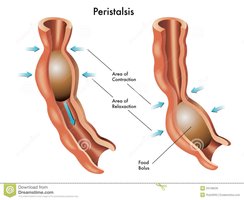
Sliding Filament Model
Muscle Contraction and Sarcomere Shortening
The sliding filament model explains how muscle contraction occurs as thin filaments slide past thick filaments, shortening the sarcomere and thus the muscle fiber. This process does not change the length of the filaments themselves, only their relative positions.
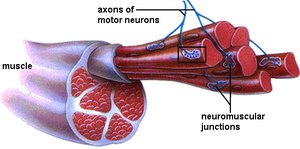
Motor Units
Organization and Recruitment
A motor unit consists of a motor neuron and all the muscle fibers it innervates. Muscles are composed of many motor units, which can be recruited in varying numbers to produce different strengths of contraction.
Small motor units: Few muscle fibers per neuron; allow fine control (e.g., eye muscles).
Large motor units: Many muscle fibers per neuron; generate powerful contractions (e.g., thigh muscles).
Muscle Twitch and Contraction Types
Muscle Twitch
A muscle twitch is the response of a motor unit to a single nerve impulse. It consists of three phases:
Latent period: Ca2+ is released from the SR; no tension produced yet.
Contraction: Cross-bridges are active; muscle tension increases.
Relaxation: Ca2+ is pumped back into the SR; muscle tension decreases.
Types of Contraction
Isometric contraction: Muscle tension is generated, but muscle length does not change.
Isotonic contraction: Muscle tension is generated, and the muscle changes length (shortens or lengthens).
Graded Muscle Responses
Temporal and Multiple Motor Unit Summation
Muscle contractions can be graded (vary in strength) through two main mechanisms:
Temporal (wave) summation: Increased frequency of stimulation before complete relaxation leads to greater tension (incomplete or complete tetanus).
Multiple motor unit summation (recruitment): Increasing stimulus strength recruits more motor units, increasing contraction strength.
Stimulus Type | Effect |
|---|---|
Threshold stimulus | Minimum stimulus required to recruit any motor units |
Maximal stimulus | Stimulus that recruits 100% of motor units |
Maximal contraction | Muscle tension when all motor units are recruited |
Example: Lifting a light object (e.g., paperclip) recruits few motor units; lifting a heavy object (e.g., bucket of water) recruits many motor units.
Additional info: The sliding filament model and cross bridge cycle are fundamental to understanding muscle physiology and are regulated by the nervous system and the availability of ATP and Ca2+.
