 Back
BackMuscles and Muscle Tissue: Structure, Function, and Contraction
Study Guide - Smart Notes
 Tailored notes based on your materials, expanded with key definitions, examples, and context.
Muscle Tissue Overview
Introduction to Muscle Tissue
Muscle tissue is essential for movement, posture, and various bodily functions. It transforms chemical energy (ATP) into mechanical energy, enabling force generation and movement. Nearly half of the body's mass is muscle tissue.
Types of muscle tissue: Skeletal, Cardiac, Smooth
Key characteristics: Excitability, Contractility, Extensibility, Elasticity
Main functions: Movement, posture maintenance, joint stabilization, heat generation
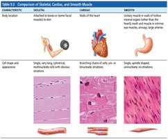
Types of Muscle Tissue
Skeletal Muscle: Attached to bones and skin, voluntary, striated, long cylindrical fibers, multinucleated, rapid contraction, tires easily.
Cardiac Muscle: Found only in the heart, involuntary, striated, branched fibers, mono- or binucleated, rhythmic contractions, does not tire easily.
Smooth Muscle: Found in walls of hollow organs (e.g., stomach, bladder), involuntary, non-striated, spindle-shaped, mononucleated, slow and sustained contractions.
Skeletal Muscle Anatomy
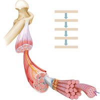
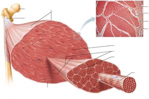
Structural Organization
Skeletal muscle is an organ composed of muscle fibers, connective tissue, blood vessels, and nerves. It is organized into several hierarchical levels:
Muscle (organ): Surrounded by epimysium
Fascicle: Bundle of muscle fibers, surrounded by perimysium
Muscle fiber (cell): Surrounded by endomysium
Myofibril: Rodlike contractile elements within muscle fibers
Myofilaments: Thick (myosin) and thin (actin) filaments
Connective Tissue Sheaths
Connective tissue sheaths support and protect muscle fibers:
Epimysium: Dense irregular connective tissue surrounding the entire muscle
Perimysium: Dense irregular connective tissue surrounding fascicles
Endomysium: Areolar connective tissue surrounding each muscle fiber
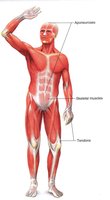
Muscle Attachments
Muscles attach to bones via tendons or aponeuroses:
Tendon: Cordlike structure attaching muscle to bone
Aponeurosis: Sheetlike structure attaching muscle to muscle or bone
Origin: Attachment to immovable bone
Insertion: Attachment to movable bone
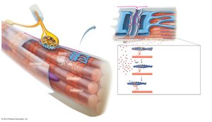
Muscle Fiber Microanatomy
Microscopic Structure
Skeletal muscle fibers are long, cylindrical cells with multiple nuclei. Key components include:
Sarcolemma: Plasma membrane of muscle fiber
Sarcoplasm: Cytoplasm containing glycosomes (glycogen storage) and myoglobin (O2 storage)
Myofibrils: Densely packed, rodlike elements responsible for muscle contraction and striations
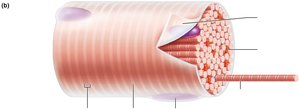
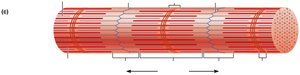
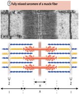
Striations and Sarcomeres
Striations are due to the arrangement of myofilaments in repeating units called sarcomeres, the functional unit of muscle contraction.
A band: Dark region with thick filaments
I band: Light region with thin filaments
Z disc: Boundary of sarcomere
H zone: Lighter region in the middle of A band
M line: Center of H zone, holds thick filaments together
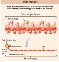
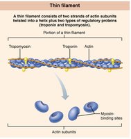
Myofilament Structure
Myofilaments are composed of contractile proteins:
Thick filaments: Made of myosin, with heads that bind actin and ATP
Thin filaments: Made of actin, with regulatory proteins troponin and tropomyosin
Muscle Contraction Mechanisms
Sliding Filament Model
Muscle contraction occurs when myosin heads bind to actin, forming cross bridges and pulling thin filaments toward the center of the sarcomere. This process shortens the muscle fiber without changing the length of the filaments.
Key steps: Cross bridge formation, power stroke, cross bridge detachment, cocking of myosin head
Excitation-Contraction Coupling
Excitation-contraction (E-C) coupling links the action potential in the sarcolemma to the sliding of myofilaments. The process involves:
Action potential travels along sarcolemma and T tubules
Ca2+ is released from the sarcoplasmic reticulum
Ca2+ binds to troponin, moving tropomyosin and exposing myosin-binding sites on actin
Myosin heads bind to actin, initiating contraction
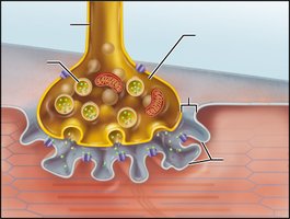
Neuromuscular Junction
The neuromuscular junction (NMJ) is the site where a motor neuron stimulates a muscle fiber. The sequence of events includes:
Action potential arrives at axon terminal
Acetylcholine (ACh) is released into the synaptic cleft
ACh binds to receptors on the sarcolemma, opening ion channels
Na+ influx causes depolarization (end plate potential)
Action potential propagates along sarcolemma
Muscle Contraction Types and Responses
Isotonic vs. Isometric Contractions
Isotonic contraction: Muscle changes length (shortens or lengthens) and moves a load
Isometric contraction: Muscle tension increases but does not change length
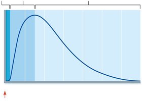
Muscle Twitch and Graded Responses
A muscle twitch is the response of a muscle to a single stimulus. It consists of three phases:
Latent period: Events of E-C coupling
Contraction period: Cross bridge formation, tension increases
Relaxation period: Ca2+ reentry into SR, tension declines
Energy for Muscle Contraction
ATP Regeneration Pathways
ATP is the immediate source of energy for muscle contraction. It is regenerated by:
Direct phosphorylation: Creatine phosphate donates phosphate to ADP
Anaerobic pathway: Glycolysis and lactic acid formation (no oxygen required)
Aerobic pathway: Cellular respiration (requires oxygen, produces most ATP)
Muscle Fiber Types and Adaptation
Classification of Muscle Fibers
Slow oxidative fibers (Type I): High endurance, aerobic, fatigue-resistant
Fast oxidative fibers (Type IIa): Intermediate properties, aerobic and anaerobic
Fast glycolytic fibers (Type IIb): Powerful, anaerobic, fatigue quickly
Adaptation to Exercise
Aerobic exercise: Increases capillaries, mitochondria, myoglobin; improves endurance
Resistance exercise: Increases muscle size (hypertrophy), strength, and connective tissue
Smooth and Cardiac Muscle
Smooth Muscle
Smooth muscle is found in the walls of hollow organs and is responsible for involuntary movements such as peristalsis. It is non-striated, spindle-shaped, and contracts slowly but can sustain contractions for long periods.
Cardiac Muscle
Cardiac muscle is found only in the heart. It is striated, branched, and connected by intercalated discs. Cardiac muscle contracts rhythmically and involuntarily, functioning as a syncytium (unit).
Clinical Terms
Convulsion: Involuntary, rapid muscle contractions
Fibrillation: Uncoordinated contraction of muscle fibers
Myalgia: Muscle pain
Muscular Dystrophy: Group of inherited diseases causing muscle degeneration
Myasthenia gravis: Autoimmune disease causing muscle weakness
Myoma: Tumor composed of muscle tissue
