 Back
BackMuscles and Muscle Tissue: Structure, Function, and Contraction
Study Guide - Smart Notes
 Tailored notes based on your materials, expanded with key definitions, examples, and context.
Muscles and Muscle Tissue
Introduction to Muscle Tissue
Muscle tissue is specialized for contraction, converting chemical energy into mechanical energy to produce force and movement. There are three main types of muscle tissue: skeletal, cardiac, and smooth muscle. Each type has unique structural and functional characteristics.
Skeletal Muscle: Voluntary, striated, attached to bones, responsible for body movement.
Cardiac Muscle: Involuntary, striated, found only in the heart, responsible for pumping blood.
Smooth Muscle: Involuntary, non-striated, found in walls of hollow organs, moves substances through internal body channels.
Skeletal Muscle Structure
Gross Anatomy of Skeletal Muscle
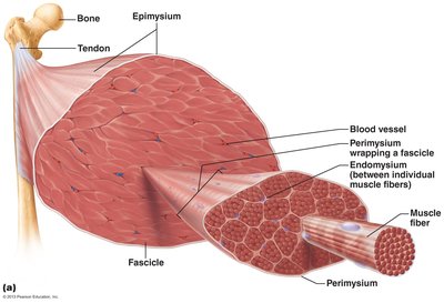
Skeletal muscle is an organ composed of muscle fibers, connective tissue, blood vessels, and nerves. It attaches to bones via tendons and is organized into bundles called fascicles.
Epimysium: Dense irregular connective tissue surrounding the entire muscle.
Perimysium: Connective tissue wrapping each fascicle (bundle of muscle fibers).
Endomysium: Fine connective tissue surrounding each individual muscle fiber.
Muscle Attachments
Muscles attach to bones at two points:
Origin: The fixed, less movable attachment.
Insertion: The movable attachment.
Attachments can be direct (muscle to bone) or indirect (via tendons or aponeuroses).
Skeletal Muscle Microscopic Anatomy
Muscle Fiber Structure
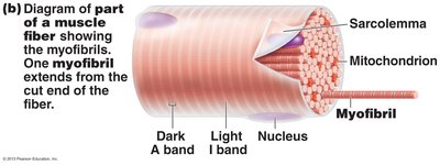
Muscle fibers are long, multinucleated cells containing specialized structures for contraction.
Sarcolemma: Plasma membrane of the muscle fiber.
Sarcoplasm: Cytoplasm containing glycosomes (glycogen storage) and myoglobin (oxygen-binding protein).
Myofibrils: Rod-like structures running the length of the cell, composed of repeating contractile units called sarcomeres.
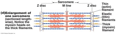
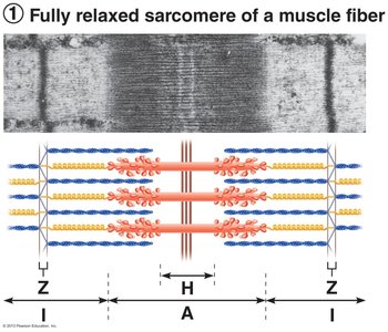
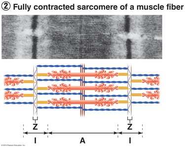
Sarcomere Structure
The sarcomere is the functional unit of muscle contraction, defined as the region between two Z discs. It contains thick (myosin) and thin (actin) filaments arranged in a precise pattern, producing the striated appearance of skeletal muscle.
A band: Dark region containing thick filaments.
I band: Light region containing thin filaments only.
H zone: Central region of A band with only thick filaments.
M line: Center of the sarcomere, holds thick filaments together.
Z disc: Boundary of the sarcomere, anchors thin filaments.
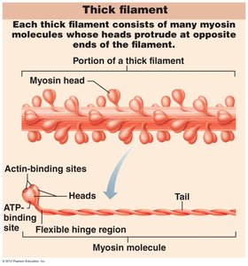
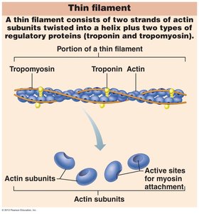
Myofilament Structure
Thick Filaments (Myosin): Composed of myosin molecules with heads that bind to actin and ATP.
Thin Filaments (Actin): Composed of actin subunits, tropomyosin, and troponin (regulatory proteins).
Muscle Contraction: The Sliding Filament Model
Mechanism of Contraction
During contraction, thin filaments slide past thick filaments, increasing the overlap between actin and myosin. The sarcomere shortens, but the filaments themselves do not change length.
Relaxed State: Minimal overlap between actin and myosin.
Contracted State: Maximum overlap, sarcomere shortens.
Neuromuscular Junction and Muscle Fiber Stimulation
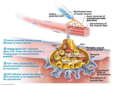
Neuromuscular Junction (NMJ)
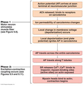
The NMJ is the site where a motor neuron communicates with a muscle fiber to initiate contraction. The process involves the release of the neurotransmitter acetylcholine (ACh), which triggers an action potential in the muscle fiber.
Action potential arrives at the axon terminal of the motor neuron.
Voltage-gated Ca2+ channels open, allowing Ca2+ to enter the axon terminal.
Ca2+ entry causes ACh to be released by exocytosis.
ACh diffuses across the synaptic cleft and binds to receptors on the sarcolemma.
Generation of Action Potential in Muscle Fiber
Binding of ACh to its receptors opens ligand-gated ion channels, allowing Na+ to enter and K+ to exit, generating an end plate potential. This local depolarization triggers an action potential that propagates along the sarcolemma and into the T-tubules, leading to muscle contraction.
Excitation-Contraction Coupling
Sequence of Events
Excitation-contraction coupling links the action potential in the sarcolemma to the activation of the myofilaments, resulting in contraction.
Action potential travels across the sarcolemma and down T-tubules.
Sarcoplasmic reticulum releases Ca2+.
Ca2+ binds to troponin, causing tropomyosin to move and expose myosin-binding sites on actin.
Myosin heads bind to actin, initiating contraction (cross-bridge cycle).
Summary Table: Types of Muscle Tissue
Feature | Skeletal Muscle | Cardiac Muscle | Smooth Muscle |
|---|---|---|---|
Location | Attached to bones | Heart walls | Walls of hollow organs |
Striations | Yes | Yes | No |
Control | Voluntary | Involuntary | Involuntary |
Cell Shape | Long, cylindrical, multinucleated | Branched, usually one nucleus | Spindle-shaped, one nucleus |
Special Features | Multiple nuclei, T-tubules, triads | Intercalated discs, pacemaker cells | Gap junctions, no T-tubules |
Key Terms and Concepts
Excitability: Ability to receive and respond to stimuli.
Contractility: Ability to shorten forcibly when stimulated.
Extensibility: Ability to be stretched or extended.
Elasticity: Ability to recoil to resting length after stretching.
Motor Unit: A motor neuron and all the muscle fibers it innervates.
Muscle Twitch: Response of a muscle to a single stimulus.
Isometric Contraction: Muscle tension increases, but muscle does not shorten.
Isotonic Contraction: Muscle changes length to move a load (concentric or eccentric).
Additional info:
ATP is required for cross-bridge cycling, Ca2+ reuptake, and Na+/K+ pump function.
Muscle metabolism involves creatine phosphate, anaerobic glycolysis, and aerobic respiration.
Muscle fiber types differ in contraction speed and metabolic pathways (slow oxidative, fast oxidative, fast glycolytic).
