 Back
BackMuscles Crossing Major Joints: Origins, Insertions, and Actions
Study Guide - Smart Notes
 Tailored notes based on your materials, expanded with key definitions, examples, and context.
Muscles Crossing the Shoulder Joint
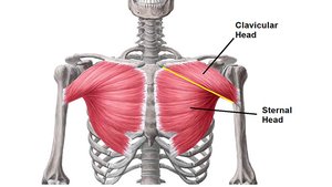
Pectoralis Major
The pectoralis major is a large, fan-shaped muscle covering the upper chest. It is essential for movements of the shoulder joint.
Origin: Clavicle, sternum, cartilage of ribs 1-6
Insertion: Intertubercular sulcus of humerus
Action: Prime mover of arm flexion; adducts and medially rotates arm
Example: Used in pushing movements such as bench press.
Deltoid
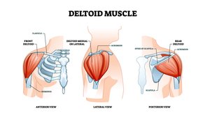
The deltoid muscle forms the rounded contour of the shoulder and is the primary abductor of the arm.
Origin: Lateral 1/3 of clavicle; acromion and spine of scapula
Insertion: Deltoid tuberosity of humerus
Action: Prime mover of arm abduction
Example: Lifting the arm sideways away from the body.
Latissimus Dorsi
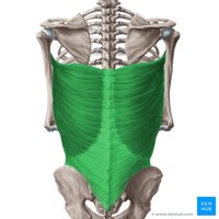
The latissimus dorsi is a broad muscle of the back, crucial for extension, adduction, and medial rotation of the arm.
Insertion: Intertubercular sulcus of humerus
Action: Prime mover of arm extension; adducts and medially rotates arm
Example: Pulling movements such as pull-ups.
Teres Major
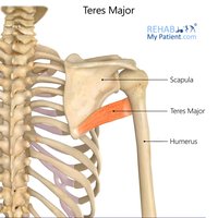
The teres major assists in movements of the shoulder, particularly extension and medial rotation.
Origin: Posterior surface at inferior angle of scapula
Insertion: Intertubercular sulcus of humerus
Action: Extends, medially rotates, and adducts arm
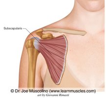
Subscapularis
The subscapularis is one of the four rotator cuff muscles, responsible for medial rotation of the arm.
Origin: Subscapular fossa of scapula
Insertion: Lesser tubercle of humerus
Action: Medially rotates arm
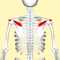
Supraspinatus
The supraspinatus initiates abduction of the arm and is part of the rotator cuff group.
Origin: Supraspinous fossa of scapula
Insertion: Greater tubercle of humerus
Action: Initiates abduction of the arm
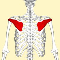
Infraspinatus
The infraspinatus is another rotator cuff muscle, important for lateral rotation of the arm.
Origin: Infraspinous fossa of scapula
Insertion: Greater tubercle of humerus
Action: Lateral rotation of arm
Teres Minor
The teres minor is a small rotator cuff muscle, also involved in lateral rotation of the arm.
Origin: Lateral border of dorsal scapular surface
Insertion: Greater tubercle of humerus
Action: Lateral rotation of arm
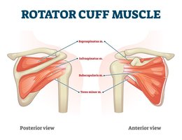
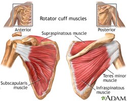
Rotator Cuff Muscles
The rotator cuff is a group of four muscles that stabilize the shoulder joint: supraspinatus, infraspinatus, teres minor, and subscapularis.
Insertion: Three of the four (supraspinatus, infraspinatus, teres minor) insert on the greater tubercle of the humerus; subscapularis inserts on the lesser tubercle.
Action: Stabilize shoulder joint; allow rotation and abduction of the arm.
Muscles Crossing the Elbow Joint
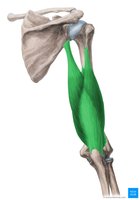
Triceps Brachii
The triceps brachii is the main extensor of the forearm at the elbow joint.
Insertion: Olecranon of ulna
Action: Powerful forearm extensor
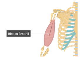
Biceps Brachii
The biceps brachii is a prominent muscle of the upper arm, responsible for flexion and supination of the forearm.
Insertion: Radial tuberosity
Action: Flexes and supinates forearm
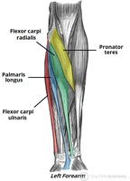
Flexors of the Forearm/Wrist
The forearm flexors originate mostly from the medial epicondyle of the humerus and are responsible for flexing the wrist and fingers.
Origin: Medial epicondyle of humerus
Action: Flexes forearm and wrist
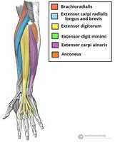
Extensors of the Forearm/Wrist
The forearm extensors originate mostly from the lateral epicondyle of the humerus and extend the wrist and fingers.
Origin: Lateral epicondyle of humerus
Action: Extends forearm and wrist
Muscles Crossing the Hip and Knee Joints
Iliopsoas
The iliopsoas is composed of the iliacus and psoas major muscles, crucial for hip flexion and trunk movement.
Action: Flex trunk at hip joint, flex thigh; lateral flexion of vertebral column
Tensor Fasciae Latae
The tensor fasciae latae helps stabilize the trunk and assists in hip abduction and medial rotation.
Action: Steadies trunk
Sartorius
The sartorius is the longest muscle in the body, crossing both the hip and knee joints.
Origin: Anterior superior iliac spine (ASIS)
Insertion: Medial tibia
Action: Flexes, abducts, and laterally rotates thigh; flexes leg
Quadriceps Femoris
The quadriceps femoris is a group of four muscles (rectus femoris, vastus lateralis, vastus medialis, vastus intermedius) responsible for knee extension.
Origin: Rectus femoris on ASIS; others on proximal femur
Insertion: Tibial tuberosity and patella
Action: Extends leg; rectus femoris also flexes thigh
Gracilis
The gracilis is a thin, superficial muscle of the medial thigh, involved in adduction and flexion.
Action: Adducts thigh; flexes and medially rotates leg
Adductors of the Thigh
The adductor group includes adductor longus, adductor brevis, adductor magnus, and gracilis, responsible for adduction, medial rotation, and flexion of the thigh.
Action: Adduct, medially rotate, and flex thigh
Gluteus Maximus
The gluteus maximus is the largest muscle of the buttocks, essential for powerful extension of the thigh.
Origin: Ilium, sacrum, and coccyx
Insertion: Gluteal tuberosity of femur
Action: Powerful thigh extensor
Gluteus Medius
The gluteus medius is located on the lateral aspect of the hip and is important for abduction and medial rotation of the thigh.
Origin: Ilium
Insertion: Greater trochanter of femur
Action: Abducts and medially rotates thigh
Gluteus Minimus
The gluteus minimus lies beneath the gluteus medius and assists in abduction and medial rotation of the thigh.
Origin: Ilium
Insertion: Greater trochanter of femur
Action: Abducts and medially rotates thigh
Hamstrings
The hamstrings are a group of three muscles (biceps femoris, semitendinosus, semimembranosus) located at the back of the thigh.
Origin: Ischial tuberosity
Action: Extends thigh; flexes leg
Muscles of the Leg
Tibialis Anterior
The tibialis anterior is the primary muscle responsible for dorsiflexion of the foot.
Action: Prime mover of dorsiflexion
Gastrocnemius
The gastrocnemius is a major calf muscle, important for plantar flexion of the foot.
Insertion: Calcaneus via calcaneal tendon
Action: Plantar flexes foot
Soleus
The soleus lies beneath the gastrocnemius and also contributes to plantar flexion.
Insertion: Calcaneus via calcaneal tendon
Action: Plantar flexes foot
Summary Table: Rotator Cuff Muscles
The following table summarizes the origins, insertions, and actions of the rotator cuff muscles:
Muscle | Origin | Insertion | Action |
|---|---|---|---|
Supraspinatus | Supraspinous fossa of scapula | Greater tubercle of humerus | Initiates abduction of arm |
Infraspinatus | Infraspinous fossa of scapula | Greater tubercle of humerus | Lateral rotation of arm |
Teres Minor | Lateral border of scapula | Greater tubercle of humerus | Lateral rotation of arm |
Subscapularis | Subscapular fossa of scapula | Lesser tubercle of humerus | Medial rotation of arm |
Additional info: The rotator cuff muscles are essential for stabilizing the shoulder joint and allowing a wide range of arm movements.
