 Back
BackMuscles of the Arms and Legs: Structure and Function
Study Guide - Smart Notes
 Tailored notes based on your materials, expanded with key definitions, examples, and context.
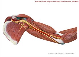
Muscles of the Scapula and Arm
Anterior View, Left Side
The anterior muscles of the scapula and arm are essential for movements such as flexion, adduction, and medial rotation of the upper limb. These muscles also stabilize the shoulder joint and facilitate complex arm movements.
Deltoid: Abducts the arm; assists in flexion and medial rotation (anterior fibers) and extension and lateral rotation (posterior fibers).
Pectoralis Major: Adducts and medially rotates the humerus; assists in flexion of the arm.
Biceps Brachii: Flexes the elbow and supinates the forearm; assists in shoulder flexion.
Brachialis: Primary flexor of the elbow.
Coracobrachialis: Flexes and adducts the arm at the shoulder.
Triceps Brachii (medial and long heads): Extends the forearm at the elbow; long head also assists in arm adduction.
Latissimus Dorsi and Teres Major: Extend, adduct, and medially rotate the arm.
Subscapularis: Medially rotates the humerus; part of the rotator cuff group.
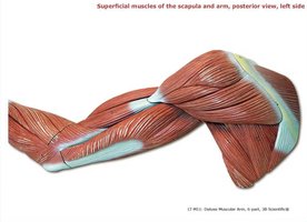
Posterior View, Left Side
The posterior muscles of the scapula and arm are primarily responsible for extension, abduction, and lateral rotation of the upper limb. They also play a key role in stabilizing the shoulder joint.
Deltoid: As above, with posterior fibers aiding in extension and lateral rotation.
Trapezius: Elevates, retracts, and rotates the scapula.
Infraspinatus and Teres Minor: Laterally rotate the arm; stabilize the shoulder joint.
Latissimus Dorsi and Teres Major: As above, extend, adduct, and medially rotate the arm.
Triceps Brachii (long and lateral heads): As above, extend the forearm at the elbow.
Brachialis and Biceps Brachii: As above, flex the elbow.
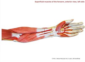
Muscles of the Forearm
Superficial Muscles, Anterior View, Left Side
The superficial muscles of the anterior forearm are primarily involved in flexion of the wrist and fingers, as well as pronation of the forearm.
Brachioradialis: Flexes the forearm at the elbow, especially when the forearm is in mid-pronation.
Flexor Carpi Radialis and Flexor Carpi Ulnaris: Flex and abduct/adduct the wrist, respectively.
Palmaris Longus: Flexes the wrist; tenses the palmar aponeurosis.
Flexor Digitorum Superficialis: Flexes the middle phalanges of the fingers.
Pronator Teres: Pronates the forearm.
Extensor Carpi Radialis Longus/Brevis, Abductor Pollicis Longus: Though primarily posterior, these muscles assist in wrist extension and thumb abduction.
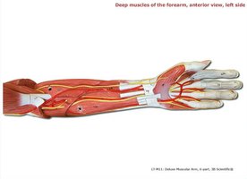
Deep Muscles, Anterior View, Left Side
The deep muscles of the anterior forearm are responsible for fine movements of the fingers and thumb, as well as pronation and supination of the forearm.
Flexor Digitorum Profundus: Flexes the distal phalanges of the fingers.
Flexor Pollicis Longus: Flexes the thumb.
Pronator Teres and Supinator: Pronate and supinate the forearm, respectively.
Palmaris Longus, Flexor Carpi Radialis, Flexor Digitorum Superficialis: As above, flex the wrist and fingers.
Abductor Pollicis Longus, Extensor Digitorum: Abduct the thumb and extend the fingers, respectively.
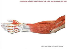
Superficial Muscles of the Forearm and Hand, Posterior View, Left Side
The posterior muscles of the forearm and hand are primarily extensors of the wrist and fingers, and abductors of the thumb and little finger.
Extensor Digitorum: Extends the fingers.
Extensor Carpi Radialis Longus/Brevis, Extensor Carpi Ulnaris: Extend and abduct/adduct the wrist.
Extensor Pollicis Longus/Brevis, Abductor Pollicis Longus: Extend and abduct the thumb.
Extensor Digiti Minimi, Extensor Indicis: Extend the little finger and index finger, respectively.
Brachioradialis, Anconeus: Assist in elbow flexion and extension.
Muscles of the Hand
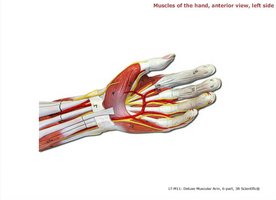
Anterior View, Left Side
The intrinsic muscles of the hand allow for fine motor movements, including flexion, extension, abduction, and adduction of the fingers and thumb.
Lumbricals: Flex the metacarpophalangeal joints and extend the interphalangeal joints.
Flexor Digitorum Profundus/Superficialis: Flex the fingers at various joints.
Flexor Pollicis Longus/Brevis, Abductor Pollicis Brevis: Flex and abduct the thumb.
Opponens Digiti Minimi, Flexor Digiti Minimi Brevis, Abductor Digiti Minimi: Control movements of the little finger.
Abductor Pollicis: Abducts the thumb.
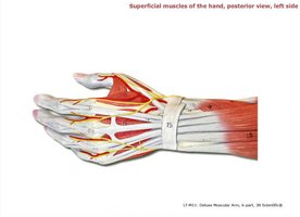
Posterior View, Left Side
The posterior hand muscles are mainly responsible for extension and abduction of the fingers and thumb, as well as adduction of the thumb.
Extensor Pollicis Longus/Brevis, Abductor Pollicis Longus: Extend and abduct the thumb.
Extensor Indicis: Extends the index finger.
Dorsal Interosseous: Abduct the fingers away from the midline of the hand.
Adductor Pollicis: Adducts the thumb toward the palm.
Abductor Digiti Minimi: Abducts the little finger.
Muscles of the Thigh
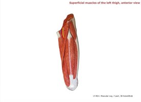
Superficial Muscles, Anterior View, Left Thigh
The anterior thigh muscles are primarily responsible for extension of the knee and flexion of the hip. They are crucial for walking, running, and maintaining posture.
Quadriceps Femoris Group (Rectus Femoris, Vastus Lateralis, Vastus Medialis, Vastus Intermedius): Extend the knee; rectus femoris also flexes the hip.
Sartorius: Flexes, abducts, and laterally rotates the thigh; flexes the knee.
Tensor Fasciae Latae: Abducts and medially rotates the thigh; stabilizes the iliotibial tract.
Adductor Longus, Pectineus, Gracilis: Adduct and flex the thigh.
Iliopsoas (Iliacus and Psoas Major): Primary hip flexor.
Deep Muscles, Anterior View, Left Thigh
The deep muscles of the anterior thigh support hip flexion and stabilization of the femur during movement.
Vastus Intermedius: Deep to rectus femoris; extends the knee.
Iliopsoas, Pectineus, Adductor Longus: As above, flex and adduct the thigh.
Sartorius (cut): As above, flexes and rotates the thigh.
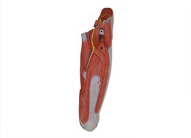
Superficial Muscles, Posterior View, Left Thigh
The posterior thigh muscles, also known as the hamstrings, are responsible for extension of the hip and flexion of the knee. They are essential for powerful lower limb movements.
Gluteus Maximus: Extends and laterally rotates the thigh.
Biceps Femoris (long and short heads), Semitendinosus, Semimembranosus: Flex the knee and extend the hip.
Adductor Magnus, Gracilis: Adduct the thigh.
Vastus Lateralis: As above, extends the knee.
Iliotibial Tract, Gluteus Medius: Stabilize the hip and thigh.
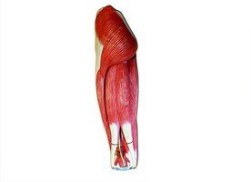
Deep Muscles, Posterior View, Left Thigh
The deep posterior thigh muscles contribute to lateral rotation, abduction, and stabilization of the hip joint.
Piriformis, Gemellus Superior/Inferior, Obturator Internus, Quadratus Femoris: Laterally rotate and stabilize the hip.
Gluteus Medius, Adductor Magnus, Gracilis: As above, abduct and adduct the thigh.
Semimembranosus, Semitendinosus (cut), Biceps Femoris (long and short heads): As above, flex the knee and extend the hip.
Popliteus: Unlocks the knee joint during flexion.
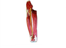
Muscles of the Leg and Foot
Superficial Muscles, Anterior View, Left Leg and Foot
The anterior compartment muscles of the leg are primarily responsible for dorsiflexion of the foot and extension of the toes.
Tibialis Anterior: Dorsiflexes and inverts the foot.
Extensor Digitorum Longus, Extensor Hallucis Longus: Extend the toes and big toe, respectively.
Fibularis Longus: Everts and plantarflexes the foot.
Superior and Inferior Extensor Retinacula: Hold tendons in place at the ankle.
Flexor Digitorum Longus, Soleus, Gastrocnemius: Plantarflex the foot and flex the toes.
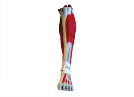
Superficial Muscles, Medial View, Left Leg and Foot
The medial view highlights muscles involved in plantarflexion, inversion, and flexion of the toes and big toe.
Tibialis Posterior: Inverts and plantarflexes the foot.
Flexor Digitorum Longus, Flexor Hallucis Longus: Flex the toes and big toe, respectively.
Abductor Hallucis, Flexor Digitorum Brevis: Abduct and flex the big toe and toes.
Calcaneal Tendon, Soleus, Gastrocnemius: As above, plantarflex the foot.
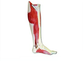
Superficial Muscles, Lateral View, Left Leg and Foot
The lateral compartment muscles are primarily responsible for eversion and plantarflexion of the foot.
Fibularis Longus, Fibularis Brevis, Fibularis Tertius: Evert the foot; longus and brevis also assist in plantarflexion.
Extensor Digitorum Brevis, Extensor Hallucis Longus: Extend the toes and big toe.
Abductor Digiti Minimi: Abducts the little toe.
Gastrocnemius, Soleus, Calcaneal Tendon: As above, plantarflex the foot.
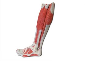
Superficial Muscles, Posterior View, Left Leg and Foot
The posterior compartment muscles are the primary plantarflexors of the foot and flexors of the toes.
Gastrocnemius, Soleus: Main plantarflexors of the foot.
Calcaneal Tendon: Common tendon for gastrocnemius and soleus, attaches to the calcaneus (heel bone).
Fibularis Longus, Fibularis Brevis: As above, evert and plantarflex the foot.
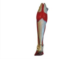
Deep Muscles, Posterior View, Left Leg and Foot
The deep posterior muscles of the leg are involved in plantarflexion, inversion, and flexion of the toes, as well as stabilization of the foot arch.
Popliteus: Unlocks the knee joint during flexion.
Tibialis Posterior: Inverts and plantarflexes the foot; supports the arch.
Flexor Digitorum Longus, Flexor Hallucis Longus: Flex the toes and big toe.
Abductor Hallucis, Flexor Digitorum Brevis, Flexor Hallucis Brevis: Abduct and flex the big toe and toes.
Lumbricals, Flexor Digiti Minimi Brevis, Abductor Digiti Minimi: Fine movements of the toes.
Plantaris: Assists in plantarflexion of the foot.
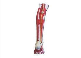
Summary Table: Major Muscle Groups of the Upper and Lower Limbs
Region | Major Muscles | Primary Actions |
|---|---|---|
Scapula & Arm (Anterior) | Deltoid, Pectoralis Major, Biceps Brachii, Brachialis | Flexion, adduction, medial rotation of arm; elbow flexion |
Scapula & Arm (Posterior) | Deltoid, Trapezius, Infraspinatus, Triceps Brachii | Extension, abduction, lateral rotation of arm; elbow extension |
Forearm (Anterior) | Flexor Carpi Radialis/Ulnaris, Palmaris Longus, Pronator Teres | Wrist/finger flexion, pronation |
Forearm (Posterior) | Extensor Digitorum, Extensor Carpi Radialis/Ulnaris | Wrist/finger extension, abduction/adduction |
Thigh (Anterior) | Quadriceps Femoris, Sartorius, Iliopsoas | Knee extension, hip flexion |
Thigh (Posterior) | Hamstrings, Gluteus Maximus | Hip extension, knee flexion |
Leg (Anterior) | Tibialis Anterior, Extensor Digitorum Longus | Dorsiflexion, toe extension |
Leg (Posterior) | Gastrocnemius, Soleus, Flexor Digitorum Longus | Plantarflexion, toe flexion |
Leg (Lateral) | Fibularis Longus/Brevis/Tertius | Eversion, plantarflexion |
