Back
BackMuscles of the Body: Head, Neck, and Trunk
Study Guide - Smart Notes
 Tailored notes based on your materials, expanded with key definitions, examples, and context.
Muscles of the Body
Overview of Axial Muscles
The axial muscles are located anterior and posterior to the body axis and are primarily responsible for movements of the trunk and maintenance of posture. These muscles include those of the thorax, abdomen, pelvis, neck, and some of the head. They play essential roles in respiration, swallowing, and supporting the vertebral column.
Muscles of the Head
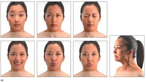
Muscles of Facial Expression
Muscles of facial expression are thin, variable in shape, and lie within the face and scalp. Unlike most skeletal muscles, they often insert into the skin rather than bones, allowing for a wide range of facial movements. These muscles are innervated by cranial nerve VII (Facial nerve).
Frontal belly of epicranius: Raises eyebrows and wrinkles the forehead.
Corrugator supercilii: Draws eyebrows together, creating a frown.
Orbicularis oculi: Closes the eyelids (blinking).
Zygomaticus major: Elevates the corners of the mouth (smiling).
Orbicularis oris: Puckers the lips (kissing).
Mentalis: Protrudes the lower lip (pouting).
Platysma: Tenses the skin of the neck.
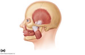
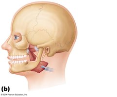
Muscles of Mastication
Mastication (chewing) involves four main pairs of muscles, all innervated by the mandibular division of the trigeminal nerve (cranial nerve V). These muscles are responsible for jaw closure, side-to-side movement, and compression of the cheeks.
Masseter and temporalis: Prime movers of jaw closure.
Pterygoid muscles: Responsible for side-to-side movements of the jaw.
Buccinator: Compresses the cheeks during chewing.
Muscles of the Neck and Throat
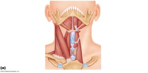
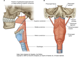
Muscles of the Anterior Neck and Throat—Swallowing
The neck is divided into anterior and posterior triangles by the sternocleidomastoid muscle. The anterior triangle contains suprahyoid and infrahyoid muscles, which are involved in swallowing. Pharyngeal constrictors squeeze food into the esophagus.
Suprahyoid muscles: Elevate the hyoid bone and floor of the mouth during swallowing.
Infrahyoid muscles: Depress the hyoid bone and larynx during swallowing and speech.
Pharyngeal constrictors: Contract sequentially to propel food into the esophagus.
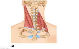
Muscles of the Neck and Vertebral Column
Movements of the Head and Neck
Muscles of the neck and vertebral column are responsible for flexion, extension, and lateral flexion of the head and neck. These muscles also help maintain posture and stabilize the head during movement.
Sternocleidomastoid: Flexes the neck and rotates the head to the opposite side.
Splenius capitis and splenius cervicis: Extend and laterally flex the head and neck.
Trapezius: Extends the neck.
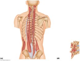
Deep Muscles of the Back (Erector Spinae Group)
The deep muscles of the back, especially the erector spinae group, are essential for trunk extension and maintaining the normal curvatures of the spine. They form a column from the sacrum to the skull and are the largest group of deep back muscles.
Erector spinae: Extends and laterally flexes the vertebral column.
Semispinalis, multifidus, rotatores: Contribute to rotation and stabilization of the vertebral column.
Muscles of the Thorax
Deep Muscles of the Thorax—Breathing
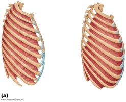
Deep muscles of the thorax are involved in the mechanics of breathing. The external intercostal muscles lift the rib cage during inspiration, while the internal intercostal muscles aid in forced expiration.
External intercostals: Elevate the ribs during inspiration.
Internal intercostals: Depress the ribs during forced expiration.
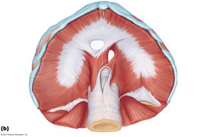
Diaphragm
The diaphragm is the most important muscle of respiration. It flattens as it contracts, increasing the volume of the thoracic cavity and allowing air to enter the lungs.
Central tendon: The insertion point for muscle fibers of the diaphragm.
Openings: For the inferior vena cava, esophagus, and aorta.
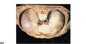
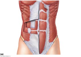
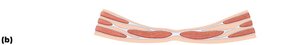
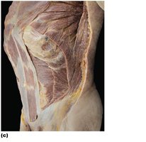
Muscles of the Abdominal Wall
Lateral and Anterior Abdominal Wall
The abdominal wall is formed by three flat muscle sheets (external oblique, internal oblique, and transversus abdominis) and a fourth muscle pair, the rectus abdominis. These muscles support the abdominal viscera, aid in trunk movement, and increase intra-abdominal pressure.
External oblique: Compresses the abdomen and flexes the vertebral column.
Internal oblique: Functions similarly to the external oblique.
Transversus abdominis: Compresses abdominal contents.
Rectus abdominis: Flexes the vertebral column and inserts at the linea alba.
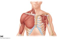
Muscles of the Thorax and Shoulder
Superficial Muscles of the Anterior Thorax
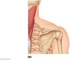
These muscles are primarily responsible for movements of the scapula and upper limb. They include the pectoralis major, pectoralis minor, serratus anterior, and subclavius.
Pectoralis major: Flexes, adducts, and medially rotates the arm.
Pectoralis minor: Draws the scapula forward and downward.
Serratus anterior: Protracts the scapula and holds it against the thoracic wall.
Subclavius: Stabilizes and depresses the clavicle.
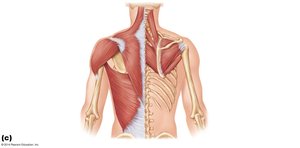
Superficial Muscles of the Posterior Thorax
These muscles move the scapula and include the trapezius, levator scapulae, rhomboid major, and rhomboid minor.
Trapezius: Elevates, retracts, and rotates the scapula.
Levator scapulae: Elevates the scapula.
Rhomboid major and minor: Retract the scapula.
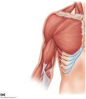
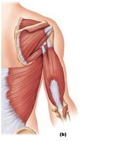
Muscles Crossing the Shoulder Joint
The shoulder joint is the most flexible joint in the body, allowing a wide range of motion but also being relatively unstable. The three largest movers of the arm at the shoulder joint are the deltoid, pectoralis major, and latissimus dorsi.
Deltoid: Abducts the arm.
Pectoralis major: Flexes, adducts, and medially rotates the arm.
Latissimus dorsi: Extends, adducts, and medially rotates the arm.