 Back
BackMuscles of the Torso and Shoulder: Structure, Function, and Clinical Relevance
Study Guide - Smart Notes
 Tailored notes based on your materials, expanded with key definitions, examples, and context.
Muscles of the Spine and Torso
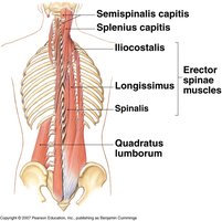
Erector Spinae Muscles
The erector spinae group is a major muscle complex responsible for maintaining posture and allowing movement of the vertebral column. It consists of three main muscle columns: iliocostalis, longissimus, and spinalis, each with subdivisions based on their location (cervicis, thoracis, lumborum).
Iliocostalis: Extends and laterally flexes the vertebral column.
Longissimus: Extends the vertebral column and head; also rotates the head.
Spinalis: Extends the vertebral column.
Quadratus lumborum: Assists in lateral flexion of the spine and stabilizes the pelvis.
Example: The erector spinae muscles are activated when standing up straight or bending backward.
Oblique and Rectus Muscles
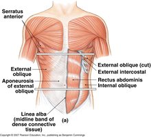
The oblique and rectus muscles form the abdominal wall, providing support for internal organs and facilitating movements such as flexion, rotation, and compression of the abdomen.
External oblique: Compresses abdomen, depresses ribs, flexes and rotates spine.
Internal oblique: Compresses abdomen, depresses ribs, flexes and rotates spine.
Rectus abdominis: Flexes vertebral column and compresses abdomen.
Serratus anterior: Protracts scapula, stabilizes shoulder.
Linea alba: Midline band of connective tissue separating left and right rectus abdominis.
Example: These muscles are engaged during sit-ups and twisting movements.
Muscles of the Shoulder and Arm
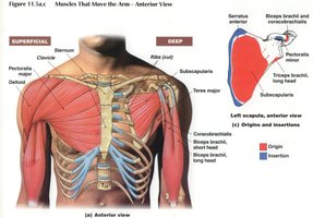
Pectoralis Major
The pectoralis major is a large, fan-shaped muscle covering the upper chest. It is crucial for movements of the shoulder joint.
Origin: Cartilage of ribs 2-6, body of sternum, inferior/medial surface of clavicle.
Insertion: Greater tubercle and intertubercular groove of humerus.
Action: Flexion, adduction, and medial rotation at the shoulder.
Innervation: Pectoral nerves.
Example: The pectoralis major is used when pushing objects or performing bench presses.
Clinical Note: Torn Pectoralis Major
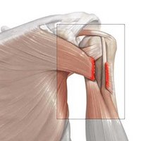
A tear in the pectoralis major muscle can result from sudden, forceful movements, often seen in athletes. Symptoms include pain, weakness, and visible deformity.
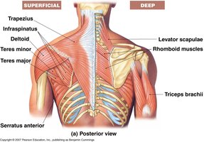
Deltoid Muscle
The deltoid muscle forms the rounded contour of the shoulder and is responsible for abduction of the arm.
Origin: Acromion and spine of scapula.
Insertion: Deltoid tuberosity of humerus.
Action: Abduction of the arm.
Innervation: Axillary nerve.
Example: Raising the arm to the side engages the deltoid muscle.
Trapezius Muscle
The trapezius is a large, triangular muscle extending from the occipital bone to the lower thoracic vertebrae and laterally to the scapula.
Origin: Occipital bone and spinous processes of C7-T12.
Insertion: Acromion and spine of scapula, lateral 1/3 of clavicle.
Action: Elevates, retracts, depresses scapula; elevates clavicle; extends neck.
Innervation: Accessory nerve.
Example: Shrugging the shoulders uses the trapezius muscle.
Latissimus Dorsi
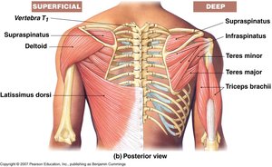
The latissimus dorsi is a broad muscle of the back, important for movements of the shoulder.
Origin: Spinous processes of T8-T12 and all lumbar vertebrae.
Insertion: Intertubercular groove of humerus.
Action: Adduction and extension of the humerus.
Innervation: Thoracodorsal nerve.
Example: Pull-ups and swimming strokes activate the latissimus dorsi.
Other Muscles of the Shoulder and Chest
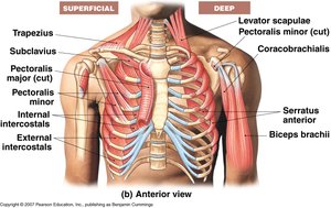
Several other muscles contribute to the movement and stabilization of the shoulder and chest.
Levator scapulae: Elevates scapula.
Rhomboid muscles: Retract scapula.
Serratus anterior: Protracts scapula.
Pectoralis minor: Depresses and protracts shoulder, depresses ribs.
Subclavius: Stabilizes clavicle.
Coracobrachialis: Flexes and adducts arm.
Biceps brachii: Flexes elbow and supinates forearm.
Summary Table: Major Muscles of the Torso and Shoulder
Muscle | Origin | Insertion | Action | Innervation |
|---|---|---|---|---|
Pectoralis Major | Ribs 2-6, sternum, clavicle | Greater tubercle, intertubercular groove of humerus | Flexion, adduction, medial rotation of shoulder | Pectoral nerves |
Deltoid | Acromion, spine of scapula | Deltoid tuberosity | Abduction of arm | Axillary nerve |
Trapezius | Occipital bone, C7-T12 | Acromion, spine of scapula, clavicle | Elevate, retract, depress scapula; extend neck | Accessory nerve |
Latissimus Dorsi | T8-T12, lumbar vertebrae | Intertubercular groove of humerus | Adduction, extension of humerus | Thoracodorsal nerve |
Oblique Muscles | Lower ribs, pelvis | Linea alba, iliac crest | Compress abdomen, flex/rotate spine | Intercostal nerves |
Key Concepts and Applications
Muscle origin and insertion determine the movement produced by contraction.
Innervation is essential for muscle function; damage to nerves can result in paralysis or weakness.
Clinical relevance: Muscle tears, such as in the pectoralis major, can significantly impair function and require medical intervention.
Additional info:
Muscles of the torso and shoulder are critical for posture, movement, and stability.
Understanding muscle anatomy aids in diagnosing injuries and planning rehabilitation.
