 Back
Muscles of the Upper Limb: Structure, Function, and Organization
Study Guide - Smart Notes
 Tailored notes based on your materials, expanded with key definitions, examples, and context.
Muscles of the Body
Pectoral Girdle and Upper Limb
The muscles of the pectoral girdle and upper limb are responsible for a wide range of movements, including flexion, extension, abduction, adduction, and rotation of the arm, forearm, wrist, and fingers. These muscles are organized into compartments based on their location and function.
Pectoral girdle muscles stabilize and move the scapula and clavicle.
Upper limb muscles are divided into those that move the arm, forearm, and hand.
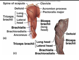
Muscles Crossing the Elbow Joint
Anterior and Posterior Compartments
The elbow joint is crossed by several muscles that allow for flexion and extension of the forearm. These muscles are grouped into anterior (flexors) and posterior (extensors) compartments.
Posterior muscles (Extensors): Triceps brachii (primary extensor), Anconeus (assists in extension).
Anterior muscles (Flexors): Biceps brachii (also supinates forearm), Brachialis, Brachioradialis.
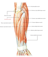
Muscles of the Forearm
Movements of the Wrist, Hand, and Fingers
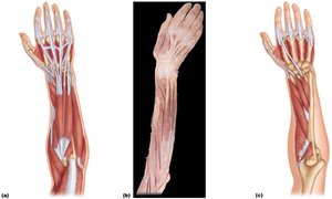
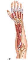
The forearm muscles control movements at the wrist and fingers. These muscles are anchored by the flexor and extensor retinacula, which hold the tendons in place. Most originate from the distal humerus and are divided into anterior (flexor) and posterior (extensor) compartments.
Movements at the wrist: Flexion, extension, abduction, and adduction.
Muscle origin: Most forearm muscles originate on the distal humerus.
Muscle action: Wrist and fingers are operated by muscles in the forearm, not the hand itself.
Anterior Flexor Compartment
The anterior compartment contains most of the digital and carpal flexors. These muscles are primarily innervated by the median and ulnar nerves and originate from a common tendon at the medial epicondyle of the humerus.
Superficial muscles: Pronator teres, flexor carpi radialis, palmaris longus, flexor carpi ulnaris, flexor digitorum superficialis.
Deep muscles: Flexor pollicis longus, flexor digitorum profundus, pronator quadratus.
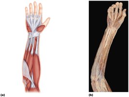
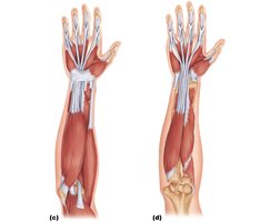
Posterior Extensor Compartment
The posterior compartment contains the extensors of the wrist and fingers. These muscles are innervated by the radial nerve and originate from a common tendon at the lateral epicondyle of the humerus.
Superficial muscles: Brachioradialis (flexes forearm), extensor carpi radialis longus, extensor carpi radialis brevis, extensor digitorum, extensor carpi ulnaris.
Deep muscles: Supinator, abductor pollicis longus, extensor pollicis brevis and longus, extensor indicis.
Summary Table: Major Muscles of the Forearm
Compartment | Muscle | Primary Action | Innervation | Origin |
|---|---|---|---|---|
Anterior (Flexor) | Flexor carpi radialis, Palmaris longus, Flexor carpi ulnaris, Flexor digitorum superficialis, Flexor pollicis longus, Flexor digitorum profundus, Pronator teres, Pronator quadratus | Flexion of wrist and fingers, pronation | Median and ulnar nerves | Medial epicondyle of humerus |
Posterior (Extensor) | Extensor carpi radialis longus, Extensor carpi radialis brevis, Extensor digitorum, Extensor carpi ulnaris, Extensor pollicis longus, Extensor pollicis brevis, Abductor pollicis longus, Extensor indicis, Supinator, Anconeus | Extension of wrist and fingers, supination | Radial nerve | Lateral epicondyle of humerus |
Key Terms and Concepts
Retinaculum: A fibrous band that holds tendons in place at the wrist.
Thenar and hypothenar muscles: Muscles at the base of the thumb (thenar) and little finger (hypothenar) that control fine movements.
Lumbricals: Small muscles in the hand that flex the metacarpophalangeal joints and extend the interphalangeal joints.
Clinical Application
Lateral epicondylitis (Tennis elbow): Inflammation of the common extensor tendon at the lateral epicondyle due to overuse.
Carpal tunnel syndrome: Compression of the median nerve under the flexor retinaculum, affecting the anterior compartment muscles.
