 Back
BackMuscles: Structure, Function, and Movement in Human Anatomy
Study Guide - Smart Notes
 Tailored notes based on your materials, expanded with key definitions, examples, and context.
Muscle Actions and Functional Groups
Overview of Muscle Actions
Muscles are responsible for producing movement by pulling on bones. They always pull a movable bone (insertion) toward a fixed bone (origin) when they contract. Muscles never push on bones. For every action performed by a muscle, there is an opposing muscle that reverses the action, known as an antagonist.
Prime Mover (Agonist): The main muscle responsible for a specific movement.
Antagonist: Muscle that opposes or reverses a particular movement.
Synergist: Assists the prime mover by adding force or reducing unnecessary movement.
Fixator: A type of synergist that stabilizes the origin of a muscle.
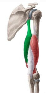
Example: The biceps brachii is the prime mover for forearm flexion, while the triceps brachii is the antagonist. For forearm extension, their roles reverse.
Naming Skeletal Muscles
Criteria for Muscle Naming
Skeletal muscles are named based on several criteria that describe their characteristics or actions:
Location: Indicates the bone or region (e.g., sternocleidomastoid).
Shape: Describes the muscle's form (e.g., deltoid means triangular).
Number of Origins: Indicates the number of attachment points (e.g., biceps has two origins, triceps has three).
Action: Describes the movement produced (e.g., flexor, extensor).
Relative Size: Terms like maximus (largest), minimus (smallest), longus (long), brevis (short).
Direction of Fibers: Rectus (straight), transversus (horizontal), oblique (diagonal).
Location of Attachments: Named for origin and insertion points.
Muscle Movements and Body Planes
Understanding Movements
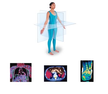
Muscle movements are described relative to anatomical position and occur in specific planes of the body:
Sagittal Plane: Divides the body into left and right parts.
Frontal (Coronal) Plane: Divides the body into anterior and posterior parts.
Transverse Plane: Divides the body into superior and inferior parts.
Angular Movements
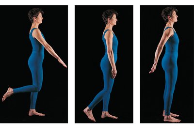
Angular movements change the angle between bones at a joint:
Flexion: Decreases the angle of the joint (e.g., bending the elbow).
Extension: Increases the angle of the joint (e.g., straightening the elbow).
Hyperextension: Extension beyond the normal range of motion.
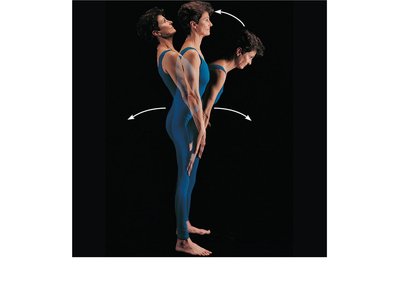
Movements in the Frontal Plane
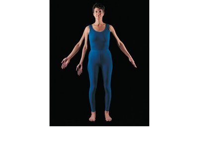
Abduction: Movement away from the midline.
Adduction: Movement toward the midline.
Circumduction: Circular movement combining flexion, abduction, extension, and adduction.
Rotational Movements
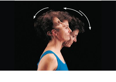
Rotation is the turning of a bone around its own long axis. Examples include shaking the head "no" (between C1 and C2 vertebrae) and rotation of the humerus or femur.
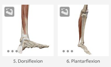
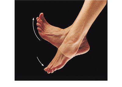
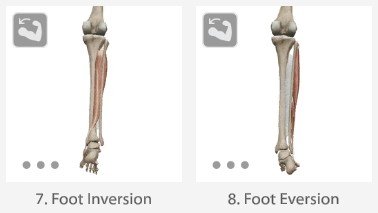
Special Movements of the Foot
Dorsiflexion: Lifting the foot so the superior surface approaches the shin.
Plantar Flexion: Pointing the toes downward.
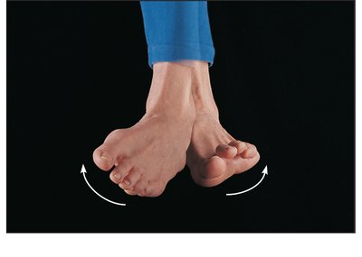
Inversion: Turning the sole of the foot medially.
Eversion: Turning the sole of the foot laterally.
Special Movements of the Mandible and Thumb
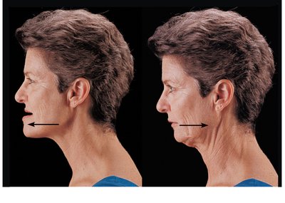
Protraction: Moving a body part anteriorly (e.g., jutting the jaw forward).
Retraction: Moving a body part posteriorly (e.g., pulling the jaw back).
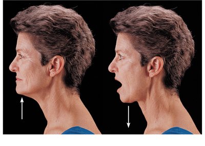
Elevation: Lifting a body part superiorly (e.g., closing the mouth).
Depression: Moving a body part inferiorly (e.g., opening the mouth).
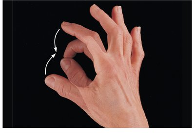
Opposition: Movement of the thumb to touch the tips of other fingers, enabling grasping.
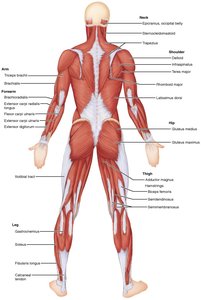
Learning Muscle Locations
Three-Dimensional Structure of Muscles
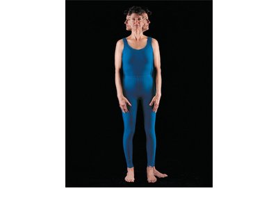
Muscles are three-dimensional organs, and many regions of the body have multiple layers of skeletal muscle. Some muscles may appear in both anterior and posterior views due to their position and orientation.
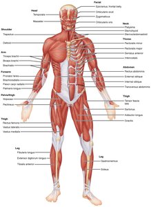
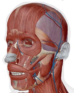
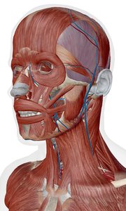
Muscles of the Head
Groups of Head Muscles
Muscles of the head are divided into two main groups:
Muscles of facial expression
Muscles of mastication (chewing) and tongue movement
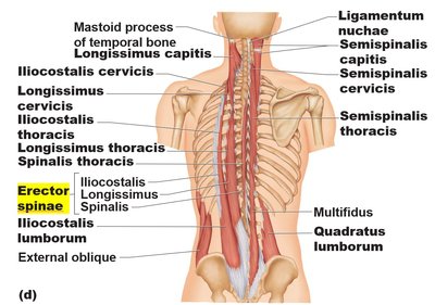
Muscles of the Neck and Vertebral Column
Head and Neck Movement
Sternocleidomastoid: Major head flexor; when both sides contract, the neck flexes. When one side contracts, the head rotates toward the opposite shoulder.
Erector Spinae Group: Prime movers of back extension and lateral bending.
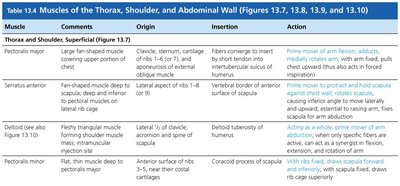
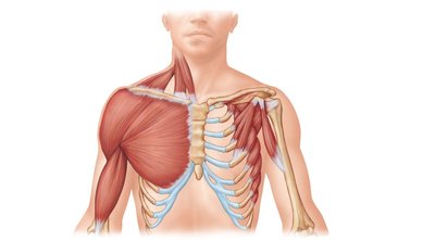
Muscles of the Thorax, Shoulder, and Abdominal Wall
Major Muscles and Their Actions
Muscle | Origin | Insertion | Action |
|---|---|---|---|
Pectoralis major | Clavicle, sternum, cartilage of ribs 1-6 (or 7), aponeurosis of external oblique muscle | Fibers converge to insert by short tendon into intertubercular sulcus of humerus | Prime mover of arm flexion; adducts, medially rotates arm |
Serratus anterior | By a series of muscle slips from ribs 1-8 (or 9) | Entire anterior surface of vertebral border of scapula | Rotates scapula so its inferior angle moves laterally and upward |
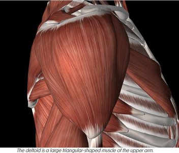
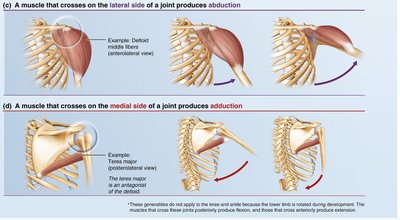
Deltoid | Lateral 1/3 of clavicle; acromion and spine of scapula | Deltoid tuberosity of humerus | Prime mover of arm abduction |
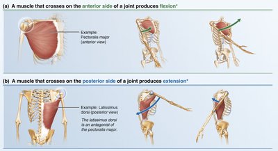
Summary Table: Muscle Actions Relative to Joint Position
Muscle Position | Movement Produced | Example |
|---|---|---|
Anterior side of joint | Flexion | Pectoralis major |
Posterior side of joint | Extension | Latissimus dorsi |
Lateral side of joint | Abduction | Deltoid middle fibers |
Medial side of joint | Adduction | Teres major |


Additional info: The above tables and images provide a comprehensive overview of muscle actions, naming conventions, and anatomical relationships, which are essential for understanding human movement and anatomy at the college level.
