 Back
BackMuscular and Nervous Tissue: Structure, Function, and Integration in the Human Body
Study Guide - Smart Notes
 Tailored notes based on your materials, expanded with key definitions, examples, and context.
Muscular Tissue
Overview and Functions
Muscular tissue is specialized for contraction, enabling movement of the body and its parts, maintenance of posture, and production of heat. There are three main types: skeletal, cardiac, and smooth muscle, each with unique structural and functional characteristics.
Skeletal muscle: Voluntary, striated, multinucleated; responsible for body movement.
Cardiac muscle: Involuntary, striated, branched; found only in the heart, responsible for pumping blood.
Smooth muscle: Involuntary, non-striated; found in walls of hollow organs, responsible for movements like peristalsis.
Myoblast fusion forms multinucleated muscle fibers, allowing for coordinated contraction and repair.
Muscle fiber structure includes the sarcolemma (cell membrane), sarcoplasm (cytoplasm), myofibrils (contractile elements), and specialized organelles such as the sarcoplasmic reticulum (SR) for calcium storage.
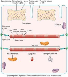
Sarcomere Structure and Function
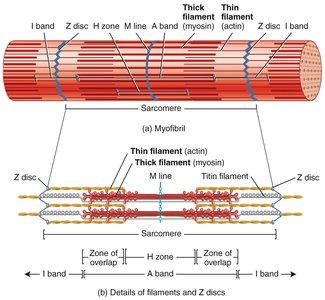
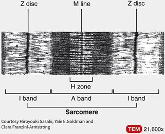
The sarcomere is the basic contractile unit of muscle fiber, defined by Z discs. It contains thick (myosin) and thin (actin) filaments, as well as regulatory proteins like troponin and tropomyosin. The arrangement of these filaments creates the striated appearance of skeletal and cardiac muscle.
Z disc: Boundary of the sarcomere; anchors thin filaments.
I band: Region with only thin filaments.
A band: Length of thick filaments; includes overlap with thin filaments.
H zone: Center of A band with only thick filaments.
M line: Center of sarcomere; holds thick filaments together.
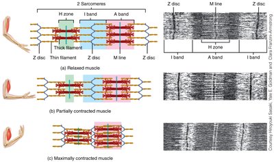
Changes During Muscle Contraction
During contraction, the sarcomere shortens as thin filaments slide past thick filaments. The I band and H zone decrease in width, while the A band remains constant.
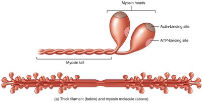
Thick and Thin Filaments
Thick filaments are composed of myosin molecules, each with a head (binding sites for actin and ATP) and a tail.
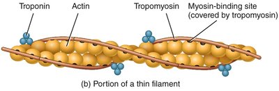
Thin filaments are primarily actin, with regulatory proteins troponin and tropomyosin controlling myosin binding.
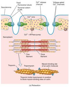
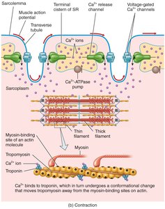
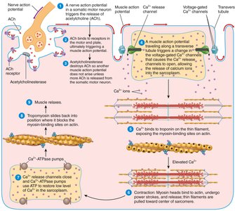
Excitation-Contraction Coupling
This process links the muscle action potential to contraction. Calcium ions released from the SR bind to troponin, causing tropomyosin to move and expose myosin-binding sites on actin, allowing cross-bridge formation and contraction.
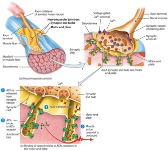
Neuromuscular Junction (NMJ)
The NMJ is the synapse between a motor neuron and a muscle fiber. An action potential in the neuron triggers acetylcholine (ACh) release, which binds to receptors on the muscle fiber, initiating a muscle action potential and contraction.
Summary of Excitation-Contraction Coupling
The sequence of events from nerve impulse to muscle contraction involves neurotransmitter release, action potential propagation, Ca2+ release, and cross-bridge cycling.
Muscle Metabolism
Muscle fibers generate ATP through three main pathways:
Creatine phosphate: Rapid ATP regeneration for short bursts.
Glycolysis: Anaerobic breakdown of glucose for moderate activity.
Mitochondrial respiration: Aerobic ATP production for sustained activity.
Oxygen debt after exercise leads to heavy breathing to restore ATP and remove lactic acid.
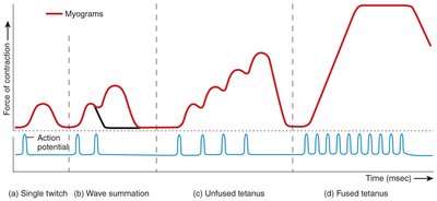
Muscle Twitch, Summation, and Tetanus
A muscle twitch is a single contraction in response to a stimulus. Wave summation occurs when stimuli are delivered before the muscle fully relaxes, increasing force. Tetanus is a sustained contraction from high-frequency stimulation.
Nervous Tissue
Overview and Functions
Nervous tissue is specialized for communication via electrical and chemical signals. It consists of neurons (excitable cells) and neuroglia (support cells). Functions include sensory input, integration, and motor output.
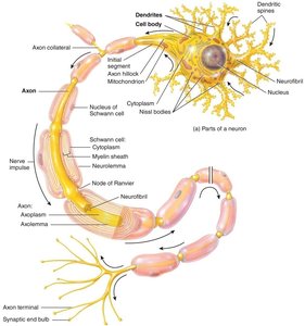
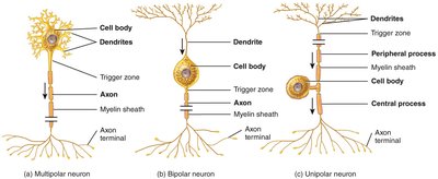
Neuron Structure and Types
Neurons have a cell body (soma), dendrites (receive signals), and an axon (transmits impulses). Nissl bodies in the soma are involved in protein synthesis. Neurons are classified by structure:
Multipolar: Many dendrites, one axon (most common in CNS).
Bipolar: One dendrite, one axon (sensory organs).
Unipolar: Single process splits into two branches (sensory neurons).
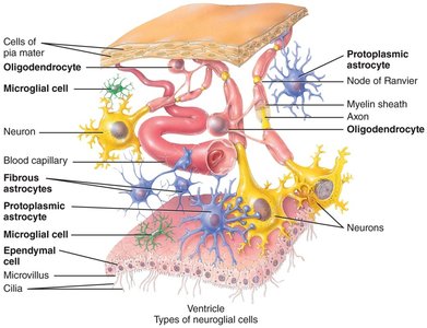
Neuroglia
Neuroglia support, protect, and nourish neurons. Types in the CNS include astrocytes, oligodendrocytes, microglia, and ependymal cells. In the PNS, Schwann cells and satellite cells are present.
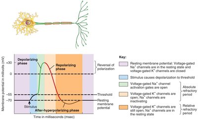
Action Potentials
Neurons generate action potentials through rapid changes in membrane potential. Key phases include depolarization (Na+ influx), repolarization (K+ efflux), and hyperpolarization. Resting membrane potential is maintained by K+ leak channels.
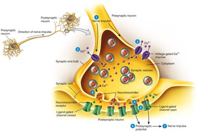
Signal Transmission at Chemical Synapses
Neurotransmitters released from presynaptic neurons bind to receptors on postsynaptic neurons, causing excitatory or inhibitory responses. Excitatory neurotransmitters depolarize the membrane, while inhibitory neurotransmitters hyperpolarize it.
CNS: Spinal Cord
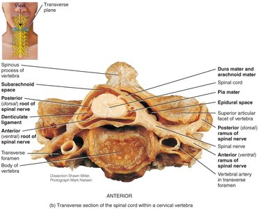
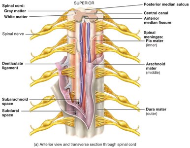
Location, Structure, and Meninges
The spinal cord is located within the vertebral foramen and is protected by three meninges: dura mater (outer), arachnoid mater (middle), and pia mater (inner). Cerebrospinal fluid (CSF) flows in the subarachnoid space.
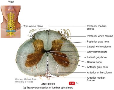
External Anatomy and Organization
The spinal cord begins at the foramen magnum and ends at L2. It is organized into gray matter (cell bodies, dendrites) and white matter (myelinated axons). Spinal nerves (31 pairs) connect the CNS to the body and are part of the PNS.
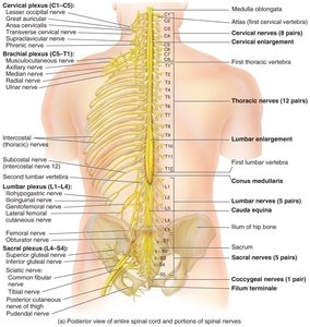
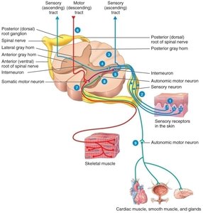
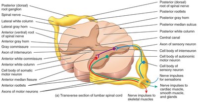
Functional Organization
Spinal nerves are mixed (sensory and motor) and named by their origin. Ascending tracts carry sensory information to the brain, while descending tracts carry motor commands. Reflex arcs involve sensory input, integration, and motor output.
Structure | Function |
|---|---|
Posterior root | Sensory input to spinal cord |
Anterior root | Motor output from spinal cord |
Gray matter | Integration of information |
White matter | Transmission of signals |
Dermatomes are skin regions supplied by specific spinal nerves, important for diagnosing nerve injuries.
CNS: The Brain
Development and Major Divisions
The brain develops from the neural tube (ectoderm). Major parts include the brain stem (medulla, pons, midbrain), diencephalon (thalamus, hypothalamus, epithalamus), cerebellum, and cerebrum (frontal, parietal, temporal, occipital, insula lobes).
Meninges and Ventricles
The brain is covered by meninges (dura mater, arachnoid mater, pia mater). The dura mater has periosteal and meningeal layers. Ventricles (lateral, third, fourth) contain CSF, which circulates through the brain and spinal cord, returning to blood via arachnoid villi.
Functional Areas and Blood Supply
Primary somatosensory area: Touch and temperature sensation (parietal lobe).
Broca’s area: Speech production (frontal lobe).
Hippocampus: Memory processing (limbic system).
Blood supply: Internal carotid and vertebral arteries, jugular vein.
Cranial Nerves and Special Functions
Optic nerve: Vision.
Vagus nerve: Innervates heart, lungs, GI tract.
Facial nerve: Motor (expression), Sensory (trigeminal).
Clinical Correlations
Alzheimer’s disease: Affects memory and cognition, especially the hippocampus.
Hemispheric lateralization: Left hemisphere for language, right for creativity.
Additional info: The notes above integrate and expand upon the provided material, ensuring a comprehensive, exam-focused review of muscular and nervous tissue, spinal cord, and brain structure and function.
