 Back
BackMuscular System: Origins, Insertions, Actions, Levers, and Naming
Study Guide - Smart Notes
 Tailored notes based on your materials, expanded with key definitions, examples, and context.
Q1. What are the definitions of origin and insertion in muscle anatomy, and how do they determine movement?
Background
Topic: Muscle Attachments and Movements
This question tests your understanding of the basic anatomical concepts of muscle origin and insertion, and how these relate to the movement of body parts.
Key Terms and Concepts:
Origin: The fixed attachment site of a muscle, usually proximal or closer to the center of the body.
Insertion: The movable attachment site, usually distal or farther from the center of the body.
Movement: When a muscle contracts, the insertion moves toward the origin, producing movement at a joint.
Step-by-Step Guidance
Recall that muscles attach to bones at two points: the origin (fixed) and the insertion (movable).
During contraction, the muscle shortens, pulling the insertion toward the origin.
Think about how this setup allows for different types of movement, such as flexion, extension, abduction, adduction, and rotation.
Consider how the specific locations of origin and insertion determine the direction and type of movement produced.
Try solving on your own before revealing the answer!
Q2. Given the origins and insertions of different muscles, what type of movement would be created when these muscles contract?
Background
Topic: Muscle Movements and Attachments
This question asks you to apply your knowledge of muscle origin and insertion to predict the movement produced by muscle contraction.
Key Terms and Concepts:
Flexion: Decreasing the angle between two bones (e.g., bending the elbow).
Extension: Increasing the angle between two bones (e.g., straightening the knee).
Abduction: Moving a limb away from the midline of the body.
Adduction: Moving a limb toward the midline of the body.
Rotation: Movement around an axis.
Step-by-Step Guidance
Identify the origin and insertion for each muscle listed (e.g., pubis to xiphoid process, manubrium to mastoid process, etc.).
Visualize or refer to anatomical diagrams to see the relative positions of these attachment points.
Consider the direction in which the insertion will move toward the origin during contraction.
Match the movement produced (flexion, extension, abduction, adduction, rotation) to the change in position caused by the muscle's contraction.
Try solving on your own before revealing the answer!
Q3. True or False: During a muscle contraction, the origin moves towards the insertion.
Background
Topic: Muscle Contraction Mechanics
This question tests your understanding of the direction of movement during muscle contraction.
Key Terms:
Origin: Fixed attachment.
Insertion: Movable attachment.
Step-by-Step Guidance
Recall the definitions of origin and insertion.
Think about which part moves during contraction (insertion moves toward origin).
Evaluate the statement and consider if it matches the anatomical reality.
Try solving on your own before revealing the answer!
Q4. The flexor carpi ulnaris attaches to both the humerus and the base of the 5th metacarpal. Which attachment is the origin?
Background
Topic: Muscle Attachments and Function
This question asks you to use the function of a muscle (flexion and adduction at the wrist) to determine which attachment is the origin.
Key Terms:
Origin: Proximal, fixed attachment.
Insertion: Distal, movable attachment.
Step-by-Step Guidance
Recall that the origin is usually closer to the trunk (proximal), and the insertion is farther away (distal).
Identify which bone (humerus or 5th metacarpal) is more proximal in the arm.
Use the direction of movement (flexion/adduction at the wrist) to confirm your answer.
Try solving on your own before revealing the answer!
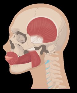
Q5. The diagram below shows some of the muscles used for chewing. Using your knowledge of the bones, identify the insertion of the masseter.
Background
Topic: Muscle Attachments in Mastication
This question tests your ability to use anatomical knowledge to identify muscle insertions based on function and location.
Key Terms:
Masseter: A major muscle involved in chewing, elevates the mandible.
Insertion: The point where the muscle attaches to the bone that moves (mandible in this case).
Step-by-Step Guidance
Recall the function of the masseter (elevates the mandible).
Identify which bone moves during chewing (mandible).
Match the muscle's action to its insertion point.
Try solving on your own before revealing the answer!
Q6. What are the roles of agonist, antagonist, synergist, and fixator muscles in movement?
Background
Topic: Muscle Actions and Coordination
This question explores the different functional roles muscles play during movement.
Key Terms:
Agonist (Prime Mover): Main muscle responsible for a movement.
Antagonist: Muscle that opposes the action of the agonist.
Synergist: Muscle that assists the agonist.
Fixator: Muscle that stabilizes the origin of the agonist.
Step-by-Step Guidance
Identify the main movement being performed (e.g., flexion, extension).
Determine which muscle is primarily responsible (agonist).
Identify the muscle that produces the opposite movement (antagonist).
Consider which muscles assist the agonist (synergists) and which stabilize the origin (fixators).
Try solving on your own before revealing the answer!
Q7. True or False: A fixator works to stabilize the insertion of the antagonist.
Background
Topic: Muscle Stabilization
This question tests your understanding of the specific role of fixator muscles during movement.
Key Terms:
Fixator: Muscle that stabilizes the origin of the agonist, not the insertion of the antagonist.
Step-by-Step Guidance
Recall the definition and function of a fixator muscle.
Consider whether the fixator acts on the origin or insertion, and whether it is related to the agonist or antagonist.
Try solving on your own before revealing the answer!
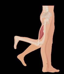
Q8. What type of lever system is the hamstring a part of when it flexes the knee, and does it work at a mechanical advantage or disadvantage?
Background
Topic: Muscle Levers and Biomechanics
This question asks you to apply your knowledge of lever systems in the body to a specific muscle action.
Key Terms and Formulas:
Lever: Rigid bar (bone) that moves on a fixed point (fulcrum, usually a joint).
Fulcrum: The pivot point (joint).
Effort: Force applied by the muscle.
Load: The weight or resistance being moved.
Mechanical Advantage: When the effort arm is longer than the load arm.
Mechanical Disadvantage: When the effort arm is shorter than the load arm.
Step-by-Step Guidance
Identify the origin and insertion of the hamstring muscles (ischial tuberosity to proximal tibia/fibula).
Visualize the movement: flexion at the knee joint.
Determine the positions of the fulcrum (knee joint), effort (hamstring attachment), and load (lower leg/foot).
Classify the lever type (first, second, or third class) based on the arrangement of fulcrum, effort, and load.
Try solving on your own before revealing the answer!
Q9. In what type of lever is the force applied between the fulcrum and the load?
Background
Topic: Lever Classes in the Human Body
This question tests your understanding of the three classes of levers and their anatomical examples.
Key Terms:
First-Class Lever: Fulcrum is between effort and load.
Second-Class Lever: Load is between fulcrum and effort.
Third-Class Lever: Effort is between fulcrum and load.
Step-by-Step Guidance
Recall the definitions and arrangements of the three lever classes.
Identify which class has the effort applied between the fulcrum and the load.
Think of anatomical examples (e.g., biceps curl for third-class lever).
Try solving on your own before revealing the answer!
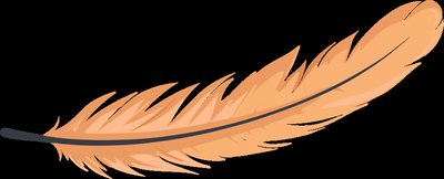
Q10. Which fascicle arrangement is most well suited to a muscle that needs to generate a lot of force over a small distance?
Background
Topic: Muscle Fascicle Arrangements
This question tests your understanding of how the arrangement of muscle fibers affects force and range of motion.
Key Terms:
Fascicle: Bundle of muscle fibers.
Pennate: Short fibers attached at an angle to a central tendon, increasing force.
Multipennate: Multiple rows of fibers attached to a central tendon, maximizing force.
Step-by-Step Guidance
Recall the different fascicle arrangements (parallel, fusiform, circular, pennate, bipennate, multipennate).
Consider which arrangement allows for the greatest number of fibers in a given area (pennate types).
Think about how fiber arrangement affects the force and range of contraction.
Try solving on your own before revealing the answer!
Q11. Based on the name, how does the fibularis brevis compare to the fibularis longus?
Background
Topic: Muscle Naming Conventions
This question tests your ability to interpret muscle names based on Latin/Greek roots and anatomical terminology.
Key Terms:
Brevis: Short.
Longus: Long.
Step-by-Step Guidance
Break down the muscle names into their Latin/Greek roots.
Compare the terms "brevis" and "longus" to determine the relative length of each muscle.
Try solving on your own before revealing the answer!
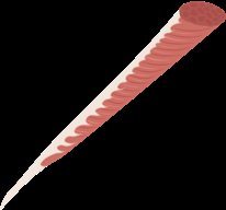
Q12. Based on the name, how would you expect the orbicularis oris muscle to be shaped?
Background
Topic: Muscle Naming and Shape
This question asks you to use the name of a muscle to predict its shape.
Key Terms:
Orbicularis: Circular.
Oris: Of the mouth.
Step-by-Step Guidance
Analyze the root "orbicularis" to determine the shape (circular).
Relate the name to the muscle's function and location (around the mouth).
Try solving on your own before revealing the answer!
