 Back
BackMuscular Tissue: Structure, Function, and Physiology
Study Guide - Smart Notes
 Tailored notes based on your materials, expanded with key definitions, examples, and context.
Muscular Tissue
Types of Muscle Tissue
Muscle tissue is specialized for contraction and is classified into three main types: skeletal, cardiac, and smooth muscle. Each type has unique structural and functional characteristics.
Skeletal Muscle: Voluntary, striated muscle attached to bones; responsible for body movement, posture, and heat production.
Cardiac Muscle: Involuntary, striated muscle found only in the heart; responsible for pumping blood.
Smooth Muscle: Involuntary, non-striated muscle found in walls of hollow organs; controls movement of substances within these organs.
Functions of Skeletal Muscle
Skeletal muscle performs several essential functions in the body:
Movement: Moves bones, enables facial expressions, speech, breathing, and swallowing.
Posture Maintenance: Stabilizes joints and maintains body position.
Protection and Support: Packages internal organs and holds them in place.
Regulation of Elimination: Circular sphincters control passage of materials at orifices.
Heat Production: Helps maintain body temperature through muscle contractions.
Characteristics of Skeletal Muscle Tissue
Skeletal muscle tissue exhibits several key physiological properties:
Excitability: Ability to respond to stimuli by changing membrane potential.
Contractility: Ability to shorten and generate force by sliding filaments.
Conductivity: Ability to propagate electrical signals along the membrane.
Extensibility: Ability to be stretched without damage.
Elasticity: Ability to return to original length after stretching or contracting.

Gross Anatomy of Skeletal Muscle
Organization and Connective Tissue Components
A skeletal muscle is an organ composed of muscle fibers, connective tissue, blood vessels, and nerves. The connective tissue layers include:
Epimysium: Dense irregular connective tissue surrounding the entire muscle.
Perimysium: Dense irregular connective tissue surrounding fascicles (bundles of muscle fibers); contains blood vessels and nerves.
Endomysium: Areolar connective tissue surrounding individual muscle fibers; provides electrical insulation and capillary support.
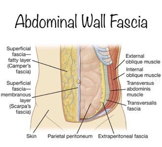
Muscle attachments can be via tendons (cordlike) or aponeuroses (sheet-like). Deep fascia separates muscles and binds those with similar functions, while superficial fascia separates muscle from skin.
Blood Supply and Innervation
Vascularization: Skeletal muscle is richly supplied with blood vessels for nutrient and waste exchange.
Innervation: Skeletal muscle is innervated by somatic motor neurons, allowing voluntary control.
Microscopic Anatomy of Skeletal Muscle
Muscle Fiber Structure
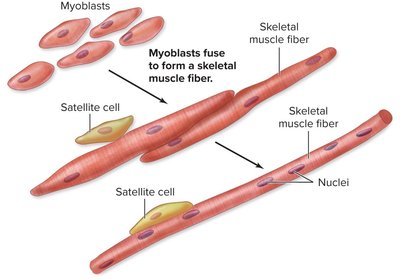
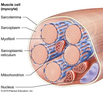
Each muscle fiber is a multinucleated cell formed by the fusion of myoblasts during development. Satellite cells remain for repair and regeneration.
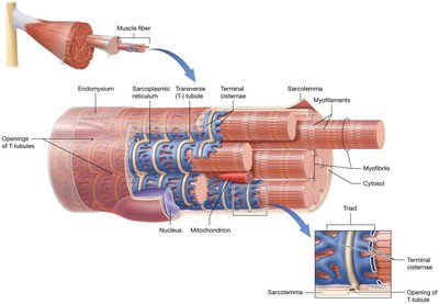
Sarcolemma: The plasma membrane of a muscle fiber, containing voltage-gated ion channels.
Sarcoplasm: The cytoplasm, containing organelles and contractile proteins.
T-tubules: Invaginations of the sarcolemma that conduct electrical signals deep into the cell.
Myofibrils: Bundles of myofilaments (contractile proteins) within the muscle fiber.
Sarcoplasmic Reticulum (SR): Specialized endoplasmic reticulum that stores and releases calcium ions.
Myofilaments and Sarcomere Organization
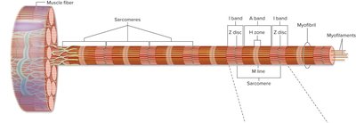
Myofibrils are composed of repeating units called sarcomeres, the functional contractile units of muscle. Sarcomeres contain thick and thin filaments:
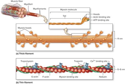
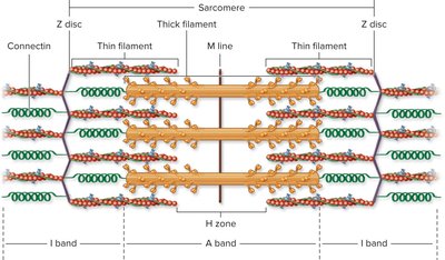
Thick Filaments: Composed of myosin molecules with heads that bind actin and hydrolyze ATP.
Thin Filaments: Composed of actin, tropomyosin, and troponin; actin has binding sites for myosin.
Regulatory Proteins: Tropomyosin and troponin regulate the interaction between actin and myosin.
Structural Proteins: Connectin (titin), dystrophin, and nebulin maintain alignment and stability.
Sarcomere Bands and Lines
Z disc: Anchors thin filaments; marks the boundary of each sarcomere.
I band: Contains only thin filaments; light region, shortens during contraction.
A band: Contains thick filaments and overlapping thin filaments; dark region.
H zone: Central region of A band with only thick filaments; disappears during maximal contraction.
M line: Center of H zone; attachment site for thick filaments.
Physiology of Skeletal Muscle Contraction
Neuromuscular Junction and Excitation-Contraction Coupling
The neuromuscular junction is the site where a motor neuron stimulates a muscle fiber. The process of muscle contraction involves:
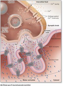
Neuromuscular Junction: Motor neuron releases acetylcholine (ACh), which binds to receptors on the motor end plate, initiating an action potential in the muscle fiber.
Excitation-Contraction Coupling: The action potential travels along the sarcolemma and T-tubules, triggering calcium release from the sarcoplasmic reticulum.
Crossbridge Cycling: Calcium binds to troponin, causing tropomyosin to move and expose binding sites on actin. Myosin heads bind to actin, perform a power stroke, release, and reset, resulting in muscle contraction.
Sliding Filament Theory
The sliding filament theory describes how thin filaments slide past thick filaments, shortening the sarcomere and producing contraction.
Muscle Relaxation
Termination of nerve signal and ACh release
Hydrolysis of ACh by acetylcholinesterase
Closure of ACh receptors and calcium channels
Return of Ca2+ to the sarcoplasmic reticulum
Return of troponin and tropomyosin to resting positions
Muscle returns to original length due to elasticity
Supplying Energy for Skeletal Muscle Metabolism
ATP Generation Pathways
Muscle fibers have limited ATP stores and rely on three main pathways for additional ATP production:
Creatine Phosphate: Provides a rapid source of phosphate to regenerate ATP from ADP.
Glycolysis: Anaerobic breakdown of glucose to produce ATP quickly but less efficiently.
Aerobic Cellular Respiration: Uses oxygen in mitochondria to produce large amounts of ATP.
Skeletal Muscle Fiber Types
Classification Criteria
Skeletal muscle fibers are classified based on contraction speed and metabolic pathway:
Slow Oxidative (Type I): Slow, fatigue-resistant, high endurance, red color due to myoglobin.
Fast Oxidative (Type IIa): Fast, intermediate endurance, primarily aerobic, light red color.
Fast Glycolytic (Type IIx): Fast, powerful, fatigable, anaerobic, white color due to low myoglobin.
Muscle Tension and Contraction Types
Muscle Twitch and Tension
A muscle twitch is a single contraction in response to a single stimulus, consisting of latent, contraction, and relaxation periods. Muscle tension is the force generated during contraction.
Isometric vs. Isotonic Contractions
Isometric: Muscle length remains constant while tension increases (e.g., holding a weight steady).
Isotonic: Muscle changes length (shortens or lengthens) while tension remains constant (e.g., lifting or lowering a weight).
Muscle Fatigue and Effects of Exercise
Muscle Fatigue
Muscle fatigue is the reduced ability to produce tension, often due to decreased glycogen stores, altered ion concentrations, or impaired excitation-contraction coupling.
Effects of Exercise and Disuse
Endurance Exercise: Increases mitochondrial density and ATP production.
Resistance Exercise: Leads to hypertrophy (increased muscle size) and some hyperplasia (increased fiber number).
Atrophy: Decrease in muscle size due to lack of use; can become permanent if prolonged.
Cardiac and Smooth Muscle
Cardiac Muscle
Cardiac muscle is found only in the heart, is striated, involuntary, and has intercalated discs for synchronized contraction.
Smooth Muscle
Smooth muscle is found in the walls of hollow organs and blood vessels. It is non-striated, involuntary, and capable of sustained contractions with low energy requirements. Smooth muscle contraction is regulated by the autonomic nervous system, hormones, and local factors.
Multiunit vs. Single-Unit Smooth Muscle
Multiunit: Each cell receives individual innervation; found in the eye and large arteries.
Single-Unit: Cells are connected by gap junctions and contract as a unit; found in most hollow organs.
