 Back
Nephrons and Renal Physiology: Structure and Function
Study Guide - Smart Notes
 Tailored notes based on your materials, expanded with key definitions, examples, and context.
Nephrons: The Functional Unit of the Kidney
Overview of Nephron Function
The nephron is the fundamental structural and functional unit of the kidney, responsible for filtering blood and forming urine. Each kidney contains over one million nephrons, which collectively process approximately 180 liters of blood-derived fluid daily, returning most to circulation and excreting only about 1% as urine.
Filtration Volume: About 120-125 mL of plasma is filtered into the renal tubules each minute.
Oxygen Consumption: Kidneys use about 10% of the body's resting oxygen supply despite being only 0.4% of body weight.
Nephron Structure: Each nephron consists of a glomerulus (capillary tuft) and a renal tubule.
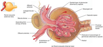
Renal Corpuscle: The glomerular (Bowman's) capsule surrounds the glomerulus, forming the renal corpuscle.
Filtration Membrane: Fenestrated endothelium allows passage of most substances except blood cells and large proteins.
Filtrate: The solute-rich fluid filtered from blood is called filtrate, which is processed into urine.
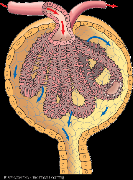
Anatomy of the Renal Tubule
The renal tubule consists of several segments, each with unique cellular features reflecting their specific roles in filtrate processing.
Proximal Convoluted Tubule (PCT): Lined with cuboidal epithelial cells with dense microvilli, increasing surface area for reabsorption.
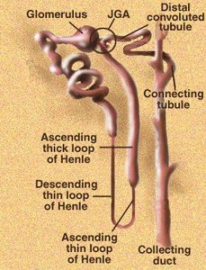
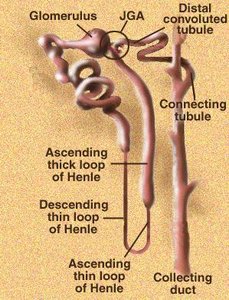
Loop of Henle: Descending limb is lined with squamous epithelium (thin segment); ascending limb transitions to cuboidal/columnar epithelium (thick segment).
Distal Convoluted Tubule (DCT): Cuboidal cells, thinner than PCT, with few microvilli.
Collecting Tubule: Receives urine from multiple nephrons, runs through medullary pyramids, and fuses into papillary ducts.
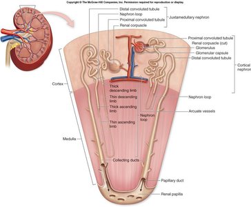
Types of Nephrons
Cortical and Juxtamedullary Nephrons
Nephrons are classified based on their location and structure, which influences their function in urine concentration.
Cortical Nephrons: Located mostly in the cortex; loops of Henle only slightly dip into the medulla.
Juxtamedullary Nephrons: Located near the cortex-medulla junction; loops of Henle deeply invade the medulla and have extensive thin segments, crucial for concentrating urine.
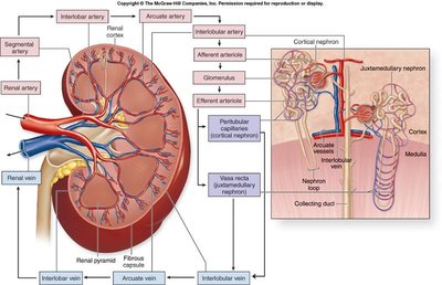
Capillary Beds of the Nephron
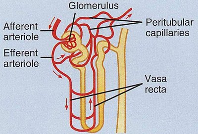
Glomerulus and Peritubular Capillaries
Each nephron is associated with two main capillary beds: the glomerulus and the peritubular capillaries, which play distinct roles in filtration and absorption.
Glomerulus: Specialized for filtration, fed and drained by afferent and efferent arterioles. High blood pressure in the glomerulus facilitates filtration.
Peritubular Capillaries: Arise from the efferent arteriole, adapted for absorption of solutes and water from the renal tubule.
Vasa Recta: Present in juxtamedullary nephrons, these vessels parallel the loop of Henle and are important for urine concentration.
Blood Pressure Gradient: Renal blood pressure drops from ~95 mmHg in arteries to ~8 mmHg in veins due to high resistance in arterioles.
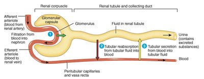
Juxtaglomerular Apparatus (JGA)
Structure and Function
The juxtaglomerular apparatus is a specialized region where the distal convoluted tubule contacts the afferent arteriole. It plays a key role in regulating blood pressure and kidney function.
Juxtaglomerular (JG) Cells: Smooth muscle cells in the afferent arteriole wall, containing renin granules; act as mechanoreceptors sensing blood pressure.
Macula Densa: Tall, closely packed DCT cells adjacent to JG cells; act as chemoreceptors/osmoreceptors responding to filtrate solute concentration.
Hormones: JGA secretes renin (regulates blood pressure) and erythropoietin (stimulates RBC production).
Glomerular Filtration
Filtration Process and Efficiency
Urine formation begins with glomerular filtration, a passive, nonselective process driven by hydrostatic pressure. The glomerulus is a highly efficient filter due to its permeability and high blood pressure.
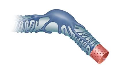
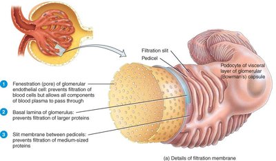
Filtration Membrane: Composed of fenestrated endothelium, basement membrane, and podocytes (visceral layer).
Efficiency: Glomerular membrane is thousands of times more permeable than other capillary beds; produces ~180L filtrate daily.
Filtration Principles: Fluid dynamics similar to tissue fluid formation elsewhere in the body.
Glomerular Filtration Rate (GFR)
The glomerular filtration rate (GFR) is a key indicator of kidney function, determined by pressures that promote or oppose filtration. GFR is regulated by renal autoregulation mechanisms and is used clinically to assess kidney health.
Normal GFR: Above 90 mL/min in healthy adults; decreases with age.
Renal Autoregulation: Feedback mechanisms adjust afferent arteriole diameter to maintain steady GFR.
Clinical Relevance: GFR is used to stage kidney disease and guide pharmacotherapy.
Estimated Creatinine Clearance Rate (eCcr): The Cockcroft-Gault formula estimates GFR:
Where Constant is 1.23 for men and 1.04 for women.
Normal Creatinine: Female: 50-90 µmol/L; Male: 70-120 µmol/L
The Filtration Membrane
Structure and Selectivity
The filtration membrane allows free passage of water and solutes smaller than plasma proteins. It consists of three layers:
Fenestrated Capillary Endothelium: Prevents passage of blood cells and large proteins.
Basement Membrane: Thin, anionic glycoproteins confer selectivity, repelling negatively charged molecules.
Podocytes (Visceral Layer): Form filtration slits that restrict passage of proteins larger than 7 nm.
Molecules smaller than 3 nm (e.g., water, glucose, amino acids, nitrogenous wastes) pass freely; larger molecules are restricted.
Colloid Osmotic Pressure: Retention of plasma proteins maintains osmotic pressure, preventing excessive water loss.
Clinical Significance: Presence of proteins or blood cells in urine indicates filtration membrane dysfunction.
Additional info: The basement membrane's negative charge favors passage of cationic and uncharged molecules, contributing to selective filtration.
