 Back
BackNervous System – Anatomy and Histology: Spinal Cord and Nerve Structure
Study Guide - Smart Notes
 Tailored notes based on your materials, expanded with key definitions, examples, and context.
Nervous System – Anatomy and Histology
Overview of Neuron Structure
The neuron is the fundamental unit of the nervous system, responsible for receiving, processing, and transmitting information through electrical and chemical signals. Neurons have specialized structures that support their function.
Dendrites: Receptive regions that receive signals from other neurons.
Cell Body (Soma): Contains the nucleus and is the biosynthetic center of the neuron.
Nucleus & Nucleolus: Control center for genetic information and ribosome production.
Axon Hillock: Region where action potentials are initiated.
Nissl Bodies: Rough endoplasmic reticulum involved in protein synthesis.
Axon: Conducts impulses away from the cell body toward other neurons or effectors.
Schwann Cells: Glial cells in the peripheral nervous system that form the myelin sheath around axons.
Myelin Sheath: Insulating layer that increases the speed of impulse conduction.
Node of Ranvier: Gaps in the myelin sheath that facilitate rapid signal transmission.
Axon Terminals: Secretory regions where neurotransmitters are released.
Types of Synapses:
Axodendritic: Between axon terminals of one neuron and dendrites of another.
Axosomatic: Between axon terminals and the cell body.
Axoaxonal: Between axon terminals of different neurons.
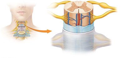
Spinal Cord and Meninges
The spinal cord is protected by three connective tissue membranes called meninges, which surround the central nervous system. These layers provide structural support and protection.
Dura Mater: The tough, outermost layer.
Arachnoid Mater: The middle, web-like layer.
Pia Mater: The delicate, innermost layer that adheres to the surface of the spinal cord.
Epidural Space: Space between the dura mater and the vertebral wall, containing fat and blood vessels.
Subdural Space: Space between the dura mater and arachnoid mater.
Subarachnoid Space: Space between the arachnoid mater and pia mater, filled with cerebrospinal fluid (CSF).
Denticulate Ligament: Extensions of pia mater that anchor the spinal cord to the dura mater.
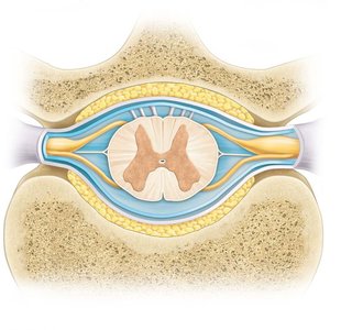
Spinal Cord Structure
The spinal cord is organized into regions of gray and white matter, and is associated with spinal nerves that connect the central nervous system to the periphery.
Spinal White Matter: Contains myelinated axons that carry information up and down the spinal cord.
Spinal Gray Matter: Contains neuron cell bodies, dendrites, and unmyelinated axons; involved in processing and integration.
Posterior Median Sulcus: Shallow groove on the dorsal side of the spinal cord.
Anterior Median Fissure: Deeper groove on the ventral side.
Nerve Roots: Bundles of axons that connect each spinal nerve to the spinal cord.
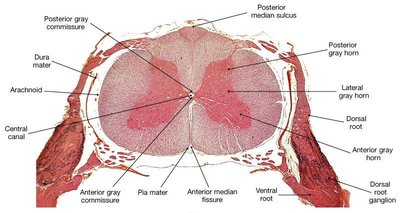
Internal Anatomy of the Spinal Cord
The spinal cord's internal structure is divided into gray and white matter, with distinct regions responsible for sensory and motor functions.
Gray Commissure: Connects the two sides of gray matter.
Posterior (Dorsal) Horn: Contains interneurons that receive sensory input.
Lateral Horn: Contains autonomic motor neurons (present in thoracic and upper lumbar regions).
Anterior (Ventral) Horn: Contains somatic motor neurons.
Central Canal: Runs through the center of the spinal cord, containing CSF.
Anterior Root (Motor): Contains motor (efferent) fibers.
Posterior Root (Sensory): Contains sensory (afferent) fibers.
Posterior Root Ganglion: Contains cell bodies of sensory neurons.
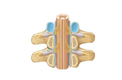
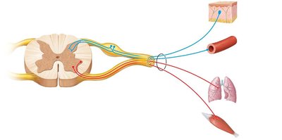
Functional Organization of Spinal Nerves
Spinal nerves are mixed nerves that carry both sensory and motor information between the spinal cord and the body. They are organized to serve specific regions and functions.
Somatic Sensory: Transmits sensory information from the skin to the CNS.
Visceral Sensory: Transmits sensory information from internal organs and blood vessels.
Somatic Motor: Controls voluntary movements of skeletal muscles.
Visceral Motor: Controls involuntary activities of smooth muscle, cardiac muscle, and glands.
Summary Table: Spinal Cord Gray Matter Regions and Functions
Region | Location | Main Function |
|---|---|---|
Posterior (Dorsal) Horn | Dorsal gray matter | Receives sensory input |
Lateral Horn | Lateral gray matter (thoracic/lumbar) | Autonomic motor neurons |
Anterior (Ventral) Horn | Ventral gray matter | Somatic motor neurons |
Gray Commissure | Central gray matter | Connects left and right sides |
Key Terms and Definitions
Neuron: Basic functional unit of the nervous system.
Myelination: Process of forming a myelin sheath around a nerve fiber.
Meninges: Three protective membranes surrounding the brain and spinal cord.
Central Canal: Fluid-filled channel in the center of the spinal cord.
Spinal Nerve: Mixed nerve carrying motor, sensory, and autonomic signals between the spinal cord and the body.
