 Back
BackNervous System Physiology: Structure, Function, and Signal Transmission
Study Guide - Smart Notes
 Tailored notes based on your materials, expanded with key definitions, examples, and context.
Nervous System Overview
Divisions of the Nervous System
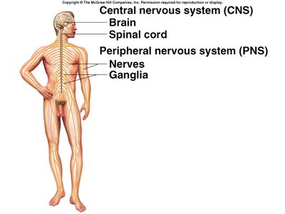
The nervous system is divided into the central nervous system (CNS) and the peripheral nervous system (PNS). The CNS consists of the brain and spinal cord, which are responsible for processing and integrating information. The PNS includes all neural tissue outside the CNS, such as nerves and ganglia, and serves as a communication network between the CNS and the rest of the body.
CNS: Brain and spinal cord; main site of information processing.
PNS: Nerves (bundles of axons) and ganglia (clusters of neuron cell bodies); transmits signals to and from the CNS.
Functional Organization of the Nervous System
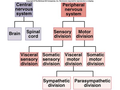
The PNS is further divided into sensory (afferent) and motor (efferent) divisions. The motor division is subdivided into the somatic and autonomic nervous systems, with the autonomic system further divided into sympathetic and parasympathetic branches.
Sensory Division: Transmits sensory information from receptors to the CNS.
Motor Division: Transmits commands from the CNS to effectors (muscles and glands).
Somatic Motor Division: Controls voluntary movements of skeletal muscles.
Autonomic Motor Division: Regulates involuntary functions (e.g., heart rate, digestion) and includes sympathetic (fight or flight) and parasympathetic (rest and digest) divisions.
Nervous System Reflex Pathways
Reflex Arc Structure and Function
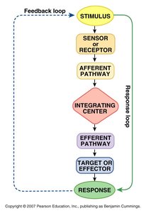
A reflex pathway is a neural circuit that mediates a reflex action. It typically involves a stimulus, sensory receptor, afferent pathway, integrating center, efferent pathway, effector, and response. Reflexes help maintain homeostasis by providing rapid, automatic responses to changes in the environment.
Stimulus: Change in the environment that initiates the reflex.
Receptor: Detects the stimulus.
Afferent Pathway: Sensory neuron transmits information to the CNS.
Integrating Center: CNS processes the information.
Efferent Pathway: Motor neuron carries command to the effector.
Effector: Muscle or gland that carries out the response.
Response: Action taken to counteract the stimulus.
Neuron Structure and Function
General Structure of Neurons
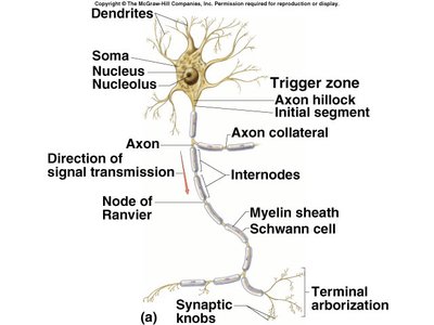
Neurons are the functional units of the nervous system, specialized for communication. They consist of a cell body (soma), dendrites, and an axon. Dendrites receive incoming signals, while the axon transmits electrical impulses away from the cell body toward other neurons or effectors.
Soma: Contains the nucleus and organelles.
Dendrites: Receive signals from other neurons.
Axon: Conducts action potentials to axon terminals.
Axon Hillock/Trigger Zone: Site where action potentials are initiated.
Myelin Sheath: Insulates axon, increasing conduction speed.
Nodes of Ranvier: Gaps in myelin sheath where action potentials are regenerated.
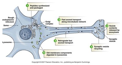
Axonal Transport
Axonal transport is the process by which materials are moved between the soma and axon terminals. It is essential for neuron function, as proteins and organelles synthesized in the soma must be delivered to the axon and synaptic terminals.
Fast Axonal Transport: Moves vesicles and organelles rapidly along microtubules using motor proteins and ATP.
Retrograde Transport: Returns used materials from axon terminal to soma for recycling.
Slow Axonal Transport: Moves cytoskeletal elements and enzymes at a slower rate.
Ion Distribution and Membrane Potentials
Ion Concentrations and Equilibrium Potentials
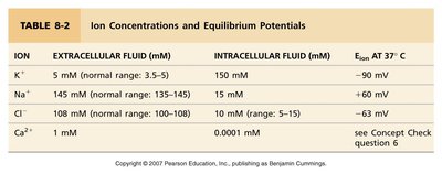
The distribution of ions across the neuronal membrane creates electrical gradients. The main ions involved are potassium (K+), sodium (Na+), chloride (Cl-), and calcium (Ca2+).
Ion | Extracellular Fluid (mM) | Intracellular Fluid (mM) | Equilibrium Potential (mV) |
|---|---|---|---|
K+ | 5 | 150 | -90 |
Na+ | 145 | 15 | +60 |
Cl- | 108 | 10 | -63 |
Ca2+ | 1 | 0.0001 | see Concept Check |
Resting Membrane Potential
The resting membrane potential is the electrical potential difference across the cell membrane when the neuron is not actively sending a signal. It is typically around -70 mV in neurons, resulting from the differential distribution of ions and selective membrane permeability, especially to potassium.
Key Factors: Ion concentration gradients and membrane permeability.
Major Contributor: Potassium leak channels allow K+ to exit the cell, making the inside more negative.
Signal Transmission: Graded and Action Potentials
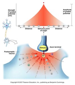
Graded Potentials
Graded potentials are small, variable changes in membrane potential that occur in the dendrites and soma. They can be depolarizing (excitatory) or hyperpolarizing (inhibitory) and decrease in strength as they spread from the site of origin.
Excitatory Postsynaptic Potentials (EPSPs): Depolarize the membrane, increasing the likelihood of an action potential.
Inhibitory Postsynaptic Potentials (IPSPs): Hyperpolarize the membrane, decreasing the likelihood of an action potential.
Summation: Multiple graded potentials can combine (spatially or temporally) to reach threshold at the trigger zone.
Action Potentials
Action potentials are large, uniform, all-or-none electrical signals that travel rapidly along the axon without losing strength. They are initiated if the membrane at the trigger zone reaches threshold (about -55 mV).
Phases: Depolarization (Na+ influx), repolarization (K+ efflux), and hyperpolarization.
Refractory Periods: Absolute (no new AP possible) and relative (stronger stimulus needed for AP).
Propagation: Action potentials regenerate at each segment of the axon, ensuring signal fidelity over long distances.
Synaptic Transmission and Neurocrines
Neurocrine Release and Types
When an action potential reaches the axon terminal, voltage-gated calcium channels open, allowing Ca2+ influx. This triggers exocytosis of synaptic vesicles, releasing neurotransmitters (neurocrines) into the synaptic cleft.
Neurotransmitters: Act rapidly at synapses (e.g., acetylcholine, glutamate, GABA).
Neuromodulators: Modulate synaptic activity, often with slower, longer-lasting effects.
Neurohormones: Released into the blood, affecting distant targets.
Neurotransmitter Receptors
Neurotransmitters bind to specific receptors on the postsynaptic cell, which may be ligand-gated ion channels (fast synaptic potentials) or G protein-coupled receptors (slow synaptic potentials).
Ligand-Gated Ion Channels: Mediate rapid EPSPs or IPSPs.
G Protein-Coupled Receptors: Activate second messenger systems for slower, prolonged responses.
Integration and Modulation of Neural Information
Summation and Modulation
Neurons integrate multiple synaptic inputs through spatial and temporal summation. Modulation can occur at both postsynaptic and presynaptic sites, allowing fine control of neural signaling.
Spatial Summation: Multiple inputs from different locations combine to reach threshold.
Temporal Summation: Rapid, successive inputs from the same source combine.
Postsynaptic Modulation: Alters the responsiveness of the postsynaptic neuron.
Presynaptic Modulation: Changes the amount of neurotransmitter released from the presynaptic neuron.
Summary Table: Key Differences Between Graded and Action Potentials
Feature | Graded Potentials | Action Potentials |
|---|---|---|
Location | Dendrites, soma | Axon |
Amplitude | Variable, graded | All-or-none, uniform |
Propagation | Decreases with distance | No loss of strength |
Summation | Possible (spatial/temporal) | Not possible (refractory period) |
Threshold | No threshold | Requires threshold (-55 mV) |
