 Back
Nervous System: Structure, Function, and Neurophysiology
Study Guide - Smart Notes
 Tailored notes based on your materials, expanded with key definitions, examples, and context.
Nervous System Overview
Introduction to the Nervous System
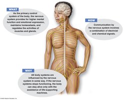
The nervous system is the primary control system of the body, responsible for regulating higher mental functions, emotional expression, homeostasis, and the activities of muscles and glands. It communicates through electrical and chemical signals, integrating sensory input and coordinating motor output.
Key Point 1: The nervous system influences all other body systems and is essential for life.
Key Point 2: Communication occurs via a combination of electrical impulses and chemical messengers.
Example: If the nervous system ceases to function, life-support machines are required to maintain bodily functions.
Nervous System Functions
Functional Components
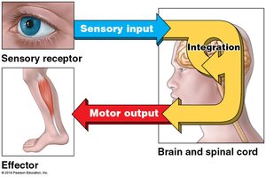
The nervous system operates through three main functions: sensory input, integration, and motor output. Sensory receptors detect changes, the brain and spinal cord integrate information, and effectors (muscles or glands) respond accordingly.
Key Point 1: Sensory input is gathered by receptors and sent to the central nervous system.
Key Point 2: Integration occurs in the brain and spinal cord, where information is processed.
Key Point 3: Motor output is the response sent to effectors to produce an action.
Example: Touching a hot object triggers sensory input, integration, and motor output (withdrawal reflex).
Homeostatic Regulation
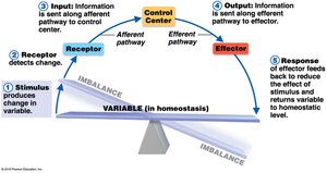
The nervous system plays a critical role in maintaining homeostasis by detecting imbalances and initiating responses to restore equilibrium.
Key Point 1: Stimulus produces a change in a variable.
Key Point 2: Receptor detects the change and sends input to the control center.
Key Point 3: Effector receives output and produces a response to restore homeostasis.
Nervous System Organization
Structural Divisions
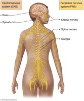
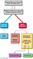
The nervous system is divided into the central nervous system (CNS) and peripheral nervous system (PNS). The CNS consists of the brain and spinal cord, while the PNS includes cranial and spinal nerves, and ganglia.
Key Point 1: CNS: Integrates and processes information.
Key Point 2: PNS: Transmits information to and from the CNS.
Example: Cranial nerves connect the brain to sensory organs; spinal nerves connect the spinal cord to the rest of the body.
Functional Divisions
The PNS is further divided into sensory (afferent) and motor (efferent) divisions. The motor division includes the somatic (voluntary) and autonomic (involuntary) systems, with the autonomic system split into sympathetic and parasympathetic branches.
Key Point 1: Sensory (afferent): Carries information from sense organs to the CNS.
Key Point 2: Motor (efferent): Carries commands from the CNS to muscles and glands.
Key Point 3: Somatic: Controls skeletal muscles; Autonomic: regulates cardiac and smooth muscle, glands.
Nervous Tissue
Structure of Nervous Tissue
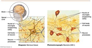
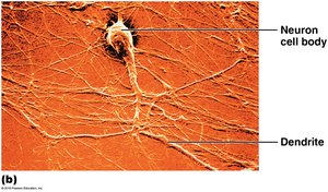
Nervous tissue is composed of neurons and supporting cells (neuroglia). Neurons are specialized for transmitting electrical impulses, while neuroglia provide structural and metabolic support.
Key Point 1: Neurons: Have a cell body, dendrites, and axon.
Key Point 2: Neuroglia: Support, protect, and nourish neurons.
Example: Photomicrographs show neuron cell bodies and processes, as well as nuclei of supporting cells.
Neuroglia
Neuroglia of the Central Nervous System (CNS)
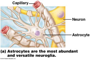
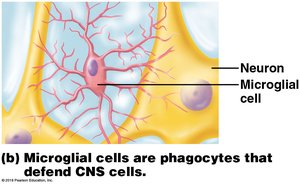
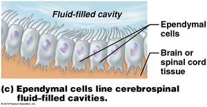
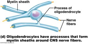
Neuroglia in the CNS include astrocytes, microglial cells, ependymal cells, and oligodendrocytes, each with distinct functions.
Key Point 1: Astrocytes: Most abundant, versatile; support neurons and maintain the blood-brain barrier.
Key Point 2: Microglial cells: Phagocytes that defend CNS cells.
Key Point 3: Ependymal cells: Line cerebrospinal fluid-filled cavities.
Key Point 4: Oligodendrocytes: Form myelin sheaths around CNS nerve fibers.
Neuroglia of the Peripheral Nervous System (PNS)
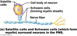
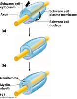
The PNS contains satellite cells and Schwann cells, which surround neurons and form myelin sheaths.
Key Point 1: Satellite cells: Surround neuron cell bodies in the PNS.
Key Point 2: Schwann cells: Form myelin sheaths around PNS nerve fibers.
Neurons
Structure of Neurons
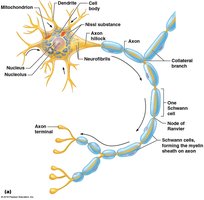
Neurons are highly specialized cells with distinct structural features, including dendrites, axons, and cell bodies. These structures facilitate the transmission of electrical impulses.
Key Point 1: Dendrites: Receive signals from other neurons.
Key Point 2: Axon: Conducts impulses away from the cell body.
Key Point 3: Cell body: Contains the nucleus and metabolic machinery.
Myelin and Conduction Velocity
Myelin Sheath and Saltatory Conduction
Myelin sheaths are insulating layers formed by Schwann cells in the PNS and oligodendrocytes in the CNS. They increase conduction velocity by enabling saltatory conduction, where impulses jump between nodes of Ranvier.
Key Point 1: Myelin: Increases speed and efficiency of electrical transmission.
Key Point 2: Saltatory conduction: Action potentials jump from node to node, rather than traveling continuously along the axon.
Structural Classification of Neurons
Types of Neurons by Structure
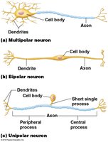
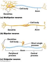
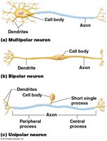
Neurons are classified as multipolar, bipolar, or unipolar based on the number and arrangement of their processes.
Key Point 1: Multipolar: Many dendrites, one axon; most common in CNS.
Key Point 2: Bipolar: One dendrite, one axon; found in sensory organs.
Key Point 3: Unipolar: Single short process; common in PNS sensory neurons.
Functional Classification of Neurons
Types of Neurons by Function
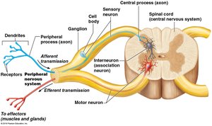
Neurons are also classified by function as sensory (afferent), motor (efferent), or interneurons (association neurons).
Key Point 1: Sensory neurons: Transmit impulses from receptors to the CNS.
Key Point 2: Motor neurons: Carry impulses from the CNS to effectors.
Key Point 3: Interneurons: Connect sensory and motor neurons within the CNS.
Types of Sensory Receptors
Classification of Sensory Receptors
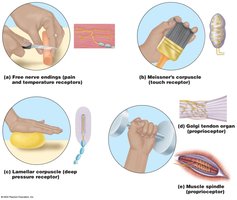
Sensory receptors are specialized to detect specific types of stimuli, such as pain, touch, pressure, and proprioception.
Key Point 1: Free nerve endings: Detect pain and temperature.
Key Point 2: Meissner's corpuscle: Touch receptor.
Key Point 3: Lamellar corpuscle: Deep pressure receptor.
Key Point 4: Golgi tendon organ and muscle spindle: Proprioceptors for muscle and tendon stretch.
Neurophysiology
Action Potential Generation and Propagation
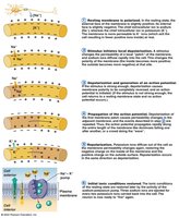
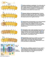
The action potential is a rapid change in membrane potential that allows neurons to transmit electrical signals. It involves depolarization, repolarization, and hyperpolarization phases.
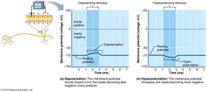
Key Point 1: Depolarization: Sodium ions (Na+) enter the cell, making the inside more positive.
Key Point 2: Repolarization: Potassium ions (K+) leave the cell, restoring negative membrane potential.
Key Point 3: Hyperpolarization: Membrane potential becomes more negative than resting.
Formula: (Membrane potential changes from resting value)
Refractory Periods
The refractory period ensures unidirectional propagation of action potentials and limits the frequency of neuronal firing.
Key Point 1: Absolute refractory period: No new action potential can be generated.
Key Point 2: Relative refractory period: A stronger stimulus is required to generate an action potential.
Synapses
Structure and Function of Synapses
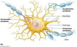
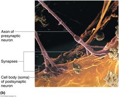
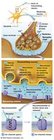
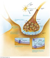
Synapses are junctions where neurons communicate with other neurons or effectors. They can be electrical or chemical, with chemical synapses being more common.
Key Point 1: Axodendritic, axosomatic, and axoaxonal synapses: Classified by location on the postsynaptic neuron.
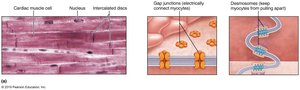
Key Point 2: Electrical synapses: Direct transfer of ions through gap junctions.
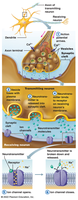
Key Point 3: Chemical synapses: Use neurotransmitters to transmit signals across a synaptic cleft.
Chemical Synapse Transmission
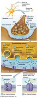
Chemical synapses transmit information via neurotransmitter release, binding, and subsequent ion channel opening in the postsynaptic cell.
Key Point 1: Neurotransmitter release: Triggered by calcium influx in the presynaptic terminal.
Key Point 2: Binding: Neurotransmitter binds to receptors, opening ion channels.
Key Point 3: Termination: Neurotransmitter effects are terminated by reuptake, degradation, or diffusion.
Major Neurotransmitters
Types and Functions of Neurotransmitters
Neurotransmitters are chemicals that transmit signals across synapses. Major types include acetylcholine, norepinephrine, dopamine, serotonin, GABA, glutamate, glycine, and endorphins.
Key Point 1: Acetylcholine: Excitatory; involved in muscle contraction.
Key Point 2: Norepinephrine: Excitatory or inhibitory; regulates mood and alertness.
Key Point 3: Dopamine: Involved in reward, motivation, and motor control.
Key Point 4: Serotonin: Regulates mood, appetite, and sleep.
Key Point 5: GABA: Main inhibitory neurotransmitter in the CNS.
Key Point 6: Glutamate: Main excitatory neurotransmitter in the CNS.
Key Point 7: Glycine: Inhibitory neurotransmitter in the spinal cord.
Key Point 8: Endorphins: Natural painkillers; modulate pain and stress.
Summary Table: Structural and Functional Classification of Neurons
Classification | Structural Types | Functional Types |
|---|---|---|
Multipolar | Many dendrites, one axon | Motor neurons, interneurons |
Bipolar | One dendrite, one axon | Sensory neurons (retina, olfactory) |
Unipolar | Single short process | Sensory neurons (PNS) |
Summary Table: Major Neuroglia Types
Cell Type | Location | Function |
|---|---|---|
Astrocyte | CNS | Support, blood-brain barrier |
Microglial cell | CNS | Phagocytosis, defense |
Ependymal cell | CNS | Line CSF cavities |
Oligodendrocyte | CNS | Myelin sheath formation |
Satellite cell | PNS | Support neuron cell bodies |
Schwann cell | PNS | Myelin sheath formation |
Additional info: Academic context was added to clarify the functions, structural classifications, and mechanisms of nervous system operation, as well as to provide self-contained explanations suitable for exam preparation.
