 Back
BackNervous Tissue and Membrane Potential: Structure, Function, and Neurophysiology
Study Guide - Smart Notes
 Tailored notes based on your materials, expanded with key definitions, examples, and context.
Nervous Tissue and Membrane Potential
Overview of Nervous Tissue
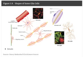
Nervous tissue is specialized for communication via electrical and chemical signals. It is composed of two main cell types: neurons, which transmit signals, and glial cells, which support and protect neurons.
Neurons: Function as the primary signaling units, analogous to electrical wires.
Glia: Provide structural and metabolic support, similar to electrical tape around wires.
Basic Structure of Neurons
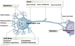
Neurons have a unique structure that enables rapid communication. They consist of three main parts:
Dendrites: Receive incoming signals from other neurons.
Cell Body (Soma): Contains the nucleus and organelles; integrates incoming signals.
Axon: Conducts electrical impulses away from the cell body toward other cells.
Special Features: Neurons are excitable, meaning they can generate and propagate electrical signals (action potentials).
Sending and Receiving Signals: The Synapse
Neurons communicate at specialized junctions called synapses. The presynaptic neuron releases neurotransmitters that cross the synaptic cleft and bind to receptors on the postsynaptic cell.
Presynaptic Cell: Sends the signal.
Postsynaptic Cell: Receives the signal.
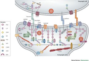
Example: Glutamate is a common excitatory neurotransmitter in the central nervous system.
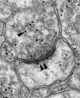
Dendritic Spines and Synaptic Plasticity
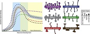
Dendritic spines are small protrusions on dendrites where synapses are often located. Their structure and number can change in response to learning and experience, a phenomenon known as synaptic plasticity.
Spine density and morphology are linked to cognitive function and neurological disorders.
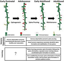
Example: Abnormal spine development is associated with Autism Spectrum Disorder (ASD).
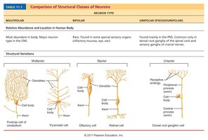
Neuron Classification
Neurons can be classified by structure and function:
Structural Types:
Multipolar: Many dendrites, one axon (most common; motor and interneurons).
Bipolar: One dendrite, one axon (special sensory organs).
Unipolar (Pseudounipolar): Single process splits into two branches (sensory neurons in PNS).
Functional Types:
Sensory (Afferent): Transmit impulses toward the CNS.
Motor (Efferent): Carry impulses away from the CNS to effectors.
Interneurons: Connect neurons within the CNS.
Neuron Type | Multipolar | Bipolar | Unipolar |
|---|---|---|---|
Relative Abundance & Location | Most abundant; major neuron type in CNS | Rare; special sensory organs (retina, olfactory) | Mainly in PNS; dorsal root ganglia |
Example | Motor neuron | Retinal cell | Sensory neuron |
Blood Brain Barrier (BBB)
The blood-brain barrier is a selective barrier that protects the brain from harmful substances while allowing essential nutrients to pass through.
Advantages: Protects neural tissue from toxins and pathogens.
Disadvantages: Limits drug delivery to the brain and can hinder treatment of CNS diseases.
The BBB is analogous to a neuron's plasma membrane: it is semi-permeable, allowing selective passage of substances.
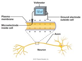
Neurophysiology: Membrane Potential
Electrical Potentials in Neurons
Neurons maintain a difference in electrical charge across their plasma membrane, known as the membrane potential. This potential is essential for the generation and propagation of nerve impulses.
Resting Membrane Potential: The voltage difference across the membrane of a resting neuron, typically around -70 mV.
Action Potential: A rapid change in membrane potential that travels along the axon.
Ion Distribution and the Na+/K+ Pump
The Na+/K+ pump is a membrane protein that actively transports sodium ions out of the cell and potassium ions into the cell, creating and maintaining the resting membrane potential.
Ions: Charged particles (e.g., Na+, K+) whose movement generates electrical currents.
Mechanism: For every 3 Na+ ions pumped out, 2 K+ ions are pumped in, resulting in a net negative charge inside the cell.
Key Equation:
where is the membrane potential.

Conceptual Analogy: Electric Potential
Electric potential in neurons can be compared to the potential for conflict in a stadium: the more fans of opposing teams and the closer they are, the greater the potential for interaction. In neurons, the greater the difference in charge and the closer the ions, the greater the membrane potential.

Example: The separation of positive and negative ions across the membrane creates the potential for electrical activity, just as separated groups of fans create the potential for interaction.
Summary Table: Key Features of Nervous Tissue and Membrane Potential
Feature | Description |
|---|---|
Neuron | Excitable cell that transmits electrical signals |
Glia | Support, protect, and nourish neurons |
Synapse | Junction for communication between neurons |
Resting Membrane Potential | Voltage difference across the membrane at rest (~-70 mV) |
Na+/K+ Pump | Maintains ion gradients and membrane potential |
Additional info: The resting membrane potential is critical for the excitability of neurons and underlies all neural signaling, including reflexes, sensation, and cognition.
