 Back
BackNervous Tissue and Nervous System: Ionic Mechanisms, Membrane Potentials, and Signal Propagation
Study Guide - Smart Notes
 Tailored notes based on your materials, expanded with key definitions, examples, and context.
Nervous Tissue and Nervous System
Ions: Sodium and Potassium
The movement of ions across neuronal membranes is fundamental to the function of the nervous system. Two key ions involved are sodium (Na+) and potassium (K+). Their distribution and movement are governed by electrochemical gradients.
Electrochemical Gradient: The combined effect of the electrical gradient (ions move toward areas of opposite charge) and the concentration gradient (ions move from areas of high to low concentration).
Direction of Ion Flow: If the gradients oppose each other, the stronger gradient determines the net flow.
Example: Potassium ions (K+) are directed by both electrical and concentration gradients across the membrane.
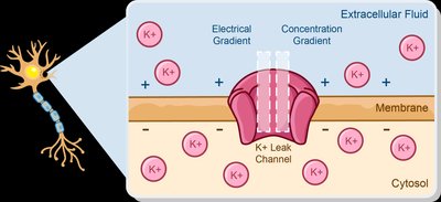
Standard Sodium and Potassium Concentrations
Neurons maintain distinct concentrations of sodium and potassium inside and outside the cell, which is essential for electrical signaling.
Sodium (Na+): Intracellular concentration is relatively low; extracellular concentration is high.
Potassium (K+): Intracellular concentration is high; extracellular concentration is low.
Example: The concentration and electrical gradients for Na+ and K+ direct their movement across the membrane.
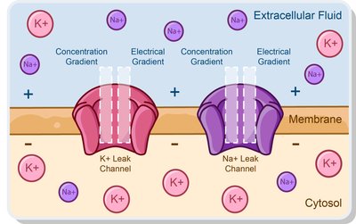
The Sodium-Potassium Pump (Na+/K+ ATPase)
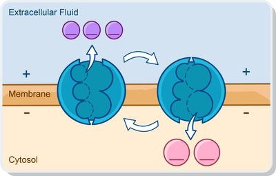
The sodium-potassium pump is an active transport mechanism that maintains the concentration gradients of Na+ and K+ across the membrane, crucial for neuronal function.
Mechanism: Uses ATP to move ions against their electrochemical gradients.
Transport: Ejects 3 Na+ ions from the cell and imports 2 K+ ions into the cell.
Importance: Maintains resting membrane potential and ionic balance.
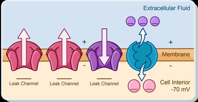
Resting Membrane Potential
The resting membrane potential is the voltage difference across the cell membrane when the neuron is not actively transmitting a signal. It is typically around -70 mV, with the cell interior more negative than the exterior.
Created by: Differences in ionic composition and membrane permeability to ions.
Stabilized by: The Na+/K+ ATPase, which maintains concentration gradients.
Change in Membrane Potential
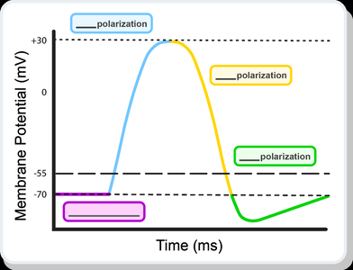
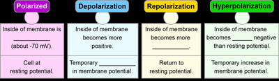
Neurons generate electrical signals by changing their membrane potential. These changes include depolarization, repolarization, and hyperpolarization.
Types of Signals: Graded potentials and action potentials.
Terminology: Polarized (resting), Depolarization (more positive), Repolarization (return to resting), Hyperpolarization (more negative than resting).
Properties of Graded and Action Potentials
Neurons use two types of electrical signals: graded potentials and action potentials. Graded potentials are variable-strength signals, while action potentials are all-or-none events.
Graded Potentials: Occur in dendrites and cell body, travel short distances, can be depolarizing or hyperpolarizing, strength depends on stimulus.
Action Potentials: Occur in axons, travel long distances, always depolarizing, initiated at threshold (~-55 mV), all-or-none phenomenon.
Graded Potentials
Graded potentials occur at the postsynaptic terminal and can be excitatory (EPSPs) or inhibitory (IPSPs).
EPSPs: Make membrane more positive (depolarized).
IPSPs: Make membrane more negative (hyperpolarized).
Sequence of Depolarizing Graded Potential: Gated Na+ channels open, Na+ enters, cell depolarizes, local current spreads, dissipates.
Summation of Graded Potentials
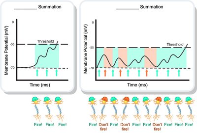
Summation is the process of adding postsynaptic potentials at the initial segment. It can be temporal (overlapping in time at one synapse) or spatial (multiple synapses in close proximity).
Action Potentials
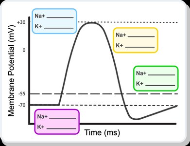
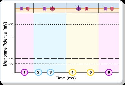
Action potentials are rapid, all-or-none electrical signals propagated along the axon. They follow a sequence of events:
Step 1: Neuron at rest
Step 2: Depolarization
Step 3: Threshold reached
Step 4: Repolarization
Step 5: Hyperpolarization
Step 6: Return to resting potential
The Refractory Period
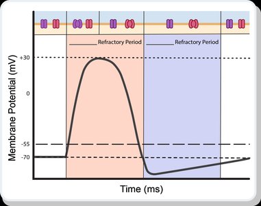
The refractory period is the time during which a neuron cannot respond to a new stimulus or requires a stronger stimulus. It ensures unidirectional propagation and limits the frequency of action potentials.
Type | Definition | Ion Channels | Time | Function |
|---|---|---|---|---|
Absolute Refractory Period | No additional action potentials can be evoked | Na+ channels open, ends when Na+ channels return to resting state | ~1 msec | Ensures distinct action potentials, unidirectional propagation |
Relative Refractory Period | Only a stronger-than-normal stimulus can evoke action potential | Na+ channels in resting state, some K+ channels open, ends when resting potential reestablished | ~2 msec | Prevents overexcitation, unidirectional propagation |
Propagation of Action Potentials
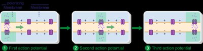
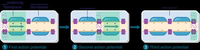
Action potentials propagate along axons by two mechanisms: continuous conduction (unmyelinated axons) and saltatory conduction (myelinated axons).
Continuous Conduction: Slow, occurs along unmyelinated axons.
Saltatory Conduction: Fast, occurs along myelinated axons, action potentials generated only at nodes of Ranvier.

Summary Table: Key Properties of Neuronal Signaling
Property | Graded Potential | Action Potential |
|---|---|---|
Type of Signal | Input signal | Output signal |
Location | Dendrites, cell body | Axon |
Travel Distance | Short | Long |
Signal Strength | Variable | All identical |
Threshold | No minimum | Initiated at threshold (~-55 mV) |
Phenomenon | Depolarizing or hyperpolarizing | Depolarizing, all-or-none |
Additional info: These notes expand on the original content by providing definitions, context, and examples for each concept, ensuring completeness and academic quality for exam preparation.
