 Back
BackNervous Tissue: Ionic Basis, Membrane Potentials, and Signal Propagation
Study Guide - Smart Notes
 Tailored notes based on your materials, expanded with key definitions, examples, and context.
Ch. 12 Nervous Tissue
Ions: Sodium and Potassium
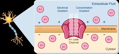
The movement of sodium (Na+) and potassium (K+) ions across the neuronal membrane is fundamental to the generation and propagation of electrical signals in nervous tissue. These movements are governed by electrochemical gradients, which combine both electrical and concentration (chemical) gradients.
Electrical Gradient: Ions move toward areas of opposite charge.
Concentration Gradient: Ions move from areas of high concentration to areas of low concentration.
Electrochemical Gradient: The net movement of ions is determined by the sum of the electrical and concentration gradients. If these gradients oppose each other, the stronger gradient determines the direction of net ion flow.
Example: For potassium ions (K+), the concentration gradient drives K+ out of the cell, while the electrical gradient pulls K+ into the cell.
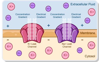
Standard Sodium and Potassium Concentrations
Neurons maintain distinct concentrations of sodium and potassium across their membranes:
Sodium (Na+): High extracellular, low intracellular concentration.
Potassium (K+): High intracellular, low extracellular concentration.
Example: At rest, Na+ is higher outside the cell, K+ is higher inside the cell.
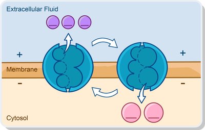
The Sodium-Potassium Pump (Na+/K+ ATPase)
The sodium-potassium pump is an active transport mechanism that maintains the resting membrane potential by moving ions against their electrochemical gradients using ATP.
Mechanism: Ejects 3 Na+ ions from the cell and imports 2 K+ ions into the cell per ATP hydrolyzed.
Importance: Maintains the concentration gradients of Na+ and K+, which are essential for electrical signaling.
Example: If the pump is blocked, intracellular K+ decreases and Na+ increases, disrupting membrane potential.
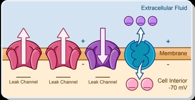
Resting Membrane Potential
The resting membrane potential is the voltage difference across the plasma membrane of a neuron when it is not actively sending a signal, typically around -70 mV. The inside of the cell is more negative than the outside.
Created by:
Differences in ionic composition of intracellular and extracellular fluids
Selective permeability of the plasma membrane to ions
Activity of the Na+/K+ ATPase
Stabilization: The sodium-potassium pump helps stabilize the resting potential by maintaining ion gradients.
Change in Membrane Potential
Types of Signals
Neurons generate two main types of electrical signals:
Graded Potentials: Small, variable-strength changes in membrane potential, usually localized.
Action Potentials: Large, uniform, all-or-none electrical impulses that propagate along the axon.
Key terminology:
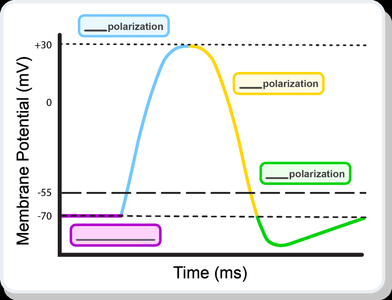
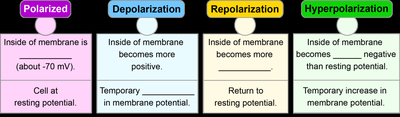
Polarization: The membrane potential is at rest (inside negative).
Depolarization: The membrane potential becomes less negative (more positive).
Repolarization: The membrane potential returns to resting value after depolarization.
Hyperpolarization: The membrane potential becomes more negative than the resting potential.
Properties of Graded and Action Potentials
Comparison of Graded and Action Potentials
Graded Potentials:
Occur in dendrites and cell body
Travel short distances
Can be depolarizing or hyperpolarizing
Magnitude depends on stimulus strength
No threshold required
Action Potentials:
Occur in axons
Travel long distances
Always depolarizing
All-or-none phenomenon (identical amplitude if threshold is reached)
Initiated at threshold (about -55 mV)
Graded Potentials
Postsynaptic Potentials
Excitatory Postsynaptic Potentials (EPSPs): Depolarize the membrane, making action potentials more likely.
Inhibitory Postsynaptic Potentials (IPSPs): Hyperpolarize the membrane, making action potentials less likely.
Sequence of a depolarizing graded potential:
Gated Na+ channels open in response to a stimulus.
Na+ enters the cell.
The inside of the cell becomes less negative (depolarizes).
Depolarization spreads locally.
The current dissipates with distance.
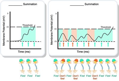
Summation of Graded Potentials
Summation is the process by which multiple graded potentials combine at the initial segment of the axon to influence whether an action potential will be generated.
Temporal Summation: Multiple graded potentials from a single synapse occur close together in time.
Spatial Summation: Graded potentials from multiple synapses occur close together in space.
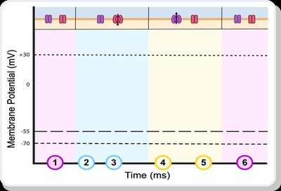
Action Potentials
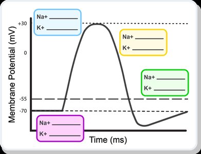
Sequence of an Action Potential
Neuron at rest
Depolarization (Na+ influx)
Threshold reached (about -55 mV)
Repolarization (K+ efflux)
Hyperpolarization
Return to resting potential
The Refractory Period
The refractory period is the time during which a neuron is less responsive to stimuli and is divided into two phases:
Absolute Refractory Period | Relative Refractory Period | |
|---|---|---|
Definition | No additional action potentials can be evoked | Only a stronger-than-normal stimulus can evoke an action potential |
Ion Channels | Na+ channels open, then inactivate | Na+ channels reset, some K+ channels open |
Function | Ensures unidirectional propagation, sets maximum firing rate | Prevents overexcitation, ensures unidirectional propagation |
Propagation of Action Potentials
Types of Propagation
Continuous Conduction: Occurs in unmyelinated axons; action potential propagates slowly along the entire membrane.
Saltatory Conduction: Occurs in myelinated axons; action potential jumps from node to node (nodes of Ranvier), increasing speed.


Summary Table: Continuous vs. Saltatory Conduction
Type | Axon Type | Speed | Where AP is Generated |
|---|---|---|---|
Continuous | Unmyelinated | Slower | Along entire axolemma |
Saltatory | Myelinated | Faster | At nodes of Ranvier |
Key Equations
Nernst Equation (for equilibrium potential of an ion): Where R = gas constant, T = temperature (K), z = charge of ion, F = Faraday's constant
Resting Membrane Potential (Goldman-Hodgkin-Katz equation): Where P = permeability of the membrane to each ion
Additional info: These notes cover the ionic basis of membrane potentials, the mechanisms of graded and action potentials, and the propagation of electrical signals in neurons, which are central to understanding nervous tissue physiology in anatomy and physiology courses.
