 Back
BackNervous Tissue: Structure, Function, and Integration CH12
Study Guide - Smart Notes
 Tailored notes based on your materials, expanded with key definitions, examples, and context.
Nervous Tissue
Overview of the Nervous System
The nervous system is responsible for internal coordination and communication within the body, working alongside the endocrine system. It utilizes neurons to transmit messages via electrical and chemical means, enabling rapid responses to stimuli.
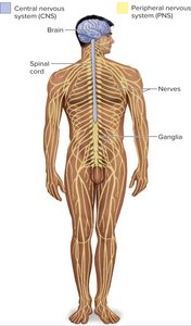
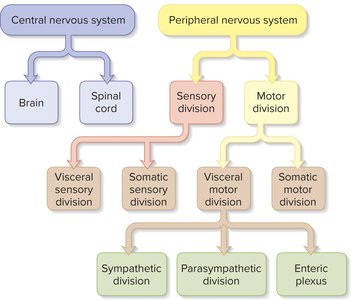
Central Nervous System (CNS): Composed of the brain and spinal cord; processes information and issues commands.
Peripheral Nervous System (PNS): Consists of nerves and ganglia; carries signals to and from the CNS.
Nerve: Bundle of axons wrapped in connective tissue.
Ganglion: Swelling in a nerve where neuron cell bodies are concentrated.
Functional Divisions of the Peripheral Nervous System
The PNS is divided into sensory and motor divisions, each with somatic and visceral subdivisions.
Sensory (afferent) division: Carries signals from receptors to CNS.
Somatic sensory division: Signals from skin, muscles, bones, joints.
Visceral sensory division: Signals from internal organs.
Motor (efferent) division: Carries signals from CNS to effectors.
Somatic motor division: Controls skeletal muscles (voluntary).
Visceral motor division (ANS): Controls glands, cardiac, and smooth muscle (involuntary).
Sympathetic division: Prepares body for action.
Parasympathetic division: Calms the body.
Enteric plexus: Coordinates digestive tract activity.
Properties and Classes of Neurons
Universal Properties of Neurons
Neurons possess three key properties that enable communication:
Excitability: Ability to respond to stimuli.
Conductivity: Ability to transmit electrical signals.
Secretion: Release of neurotransmitters at axon terminals.
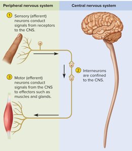
Functional Classes of Neurons
Sensory (afferent) neurons: Detect stimuli and transmit information to CNS.
Interneurons: Process information within CNS; most numerous.
Motor (efferent) neurons: Send signals from CNS to effectors (muscles/glands).
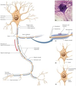
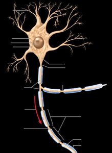
Structure of a Neuron
Principal Components
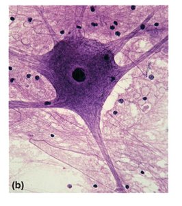
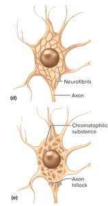
Neurons consist of a cell body (soma), dendrites, and an axon.
Cell body (soma): Contains nucleus and organelles; site of metabolic activity.
Dendrites: Branching extensions; receive signals from other neurons.
Axon: Long, cylindrical extension; transmits signals to other cells.
Axon hillock: Origin of axon; important in initiating action potentials.
Terminal arborization: Branching at axon end; forms synapses.
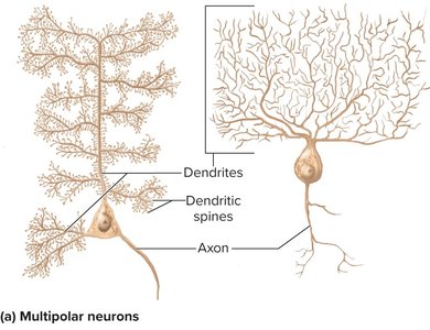
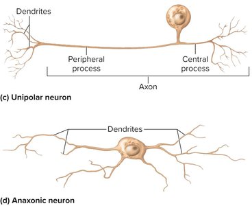
Structural Classes of Neurons
Neurons are classified by the number and arrangement of their processes:
Multipolar: One axon, multiple dendrites; most common in CNS.
Bipolar: One axon, one dendrite; found in sensory organs.
Unipolar: Single process splits into peripheral and central branches.
Anaxonic: Many dendrites, no axon; found in brain and retina.
Axonal Transport
Axonal transport is the movement of materials between the cell body and axon terminals.
Anterograde transport: Movement away from cell body; driven by kinesin.
Retrograde transport: Movement toward cell body; driven by dynein.
Fast axonal transport: 200–400 mm/day; moves organelles, vesicles.
Slow axonal transport: 0.2–0.5 mm/day; moves cytoskeletal components.
Supportive Cells (Neuroglia)
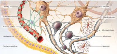
Types of Glia in CNS
Neuroglia support neurons and maintain the nervous system environment.
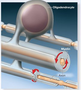
Oligodendrocytes: Form myelin sheaths in CNS.
Ependymal cells: Line brain cavities; produce cerebrospinal fluid.
Microglia: Macrophages; remove debris and defend against pathogens.
Astrocytes: Most abundant; provide structural support, regulate blood-brain barrier, supply nutrients, and form scar tissue.
Types of Glia in PNS
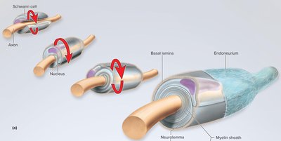
Schwann cells: Form myelin sheath in PNS; assist in nerve regeneration.
Satellite cells: Surround neuron cell bodies in ganglia; regulate environment.
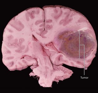
Brain Tumors
Gliomas: Tumors of glial cells; highly malignant.
Blood-brain barrier: Reduces effectiveness of chemotherapy.
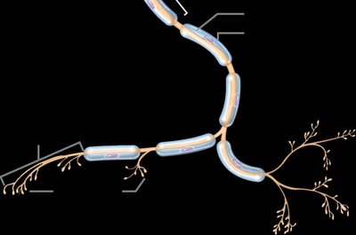
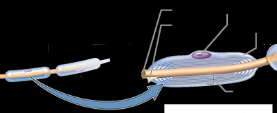
Myelin Sheath
Myelination in PNS
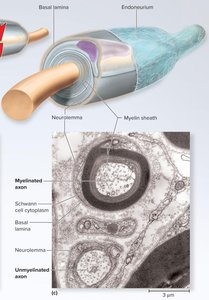
The myelin sheath is a spiral layer of insulation around axons, formed by Schwann cells in the PNS.
Neurilemma: Outermost coil containing Schwann cell nucleus.
Basal lamina and endoneurium: External layers providing support.
Myelination in CNS
Oligodendrocytes myelinate axons in the CNS by wrapping processes around multiple axons.
No neurilemma: Unlike PNS, CNS myelin lacks neurilemma.
Myelination spirals inward: New layers pushed under older ones.
Segmented Structure of Myelin
Node of Ranvier: Gap between myelin segments.
Internodal segments: Myelin-covered regions.
Initial segment: Bare axon between hillock and first glial cell.
Trigger zone: Site of action potential initiation.
Diseases of the Myelin Sheath
Multiple Sclerosis (MS): CNS myelin deteriorates; conduction disrupted.
Tay–Sachs Disease: Accumulation of glycolipid in myelin; fatal in early childhood.
Unmyelinated Axons
Structure in PNS
Schwann cells hold unmyelinated axons in surface grooves, wrapping once around each axon. 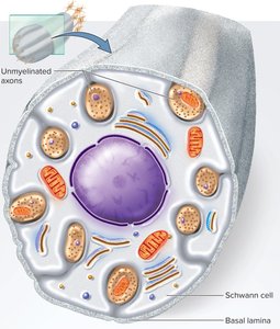
Conduction Speed of Axons
Diameter: Larger axons conduct signals faster.
Myelin: Presence of myelin increases conduction speed.
Examples: Small, unmyelinated fibers: 0.5–2.0 m/s; large, myelinated fibers: up to 120 m/s.
Nerve Regeneration
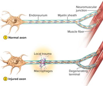
Regeneration in PNS
Damaged axons can regenerate if the cell body and neurilemma remain intact.
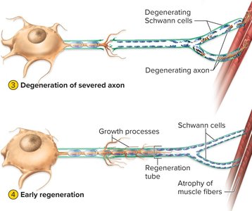
Distal axon degenerates; macrophages clean debris.
Cell body swells; ER breaks up; nucleus moves off center.
Axon stump sprouts growth processes.
Schwann cell, endoneurium, and basal lamina form regeneration tube.
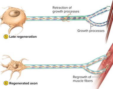
Regrowth guided to original destination.
Contact reestablished; neurosoma returns to normal; muscle fibers regrow.
Electrophysiology of Neurons
Electrical Potentials and Currents
Neural communication relies on electrical potentials and currents.
Electrical potential: Difference in concentration of charged particles; measured in volts.
Current: Flow of charged particles.
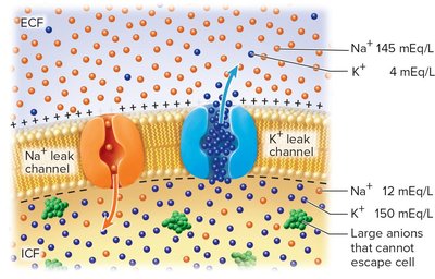
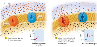
Resting membrane potential (RMP): Typically −70 mV in resting neuron.
Ionic Basis of RMP
Potassium (K+): Most influential; more concentrated inside cell.
Sodium (Na+): More concentrated outside cell; less permeable.
Sodium-potassium pump: Maintains gradients; uses ATP.
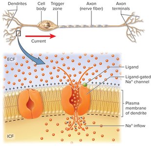
Local Potentials
Local potentials are temporary changes in membrane potential caused by stimulation.
Depolarization: Entry of Na+ makes membrane less negative.
Graded: Vary in magnitude.
Decremental: Weaken with distance.
Reversible: Return to RMP if stimulus ceases.
Excitatory or inhibitory: Depolarization (excites), hyperpolarization (inhibits).
Action Potentials
Action potentials are rapid changes in voltage due to opening and closing of voltage-gated ion channels.
Local potential reaches trigger zone.
Threshold reached (~−55 mV); Na+ channels open.
Na+ enters, depolarizes membrane to +35 mV.
K+ channels open; K+ exits, repolarizes membrane.
Hyperpolarization occurs; membrane returns to RMP.
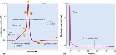
Refractory Period
Absolute refractory period: No new action potential can be triggered.
Relative refractory period: Stronger stimulus required during hyperpolarization.
Signal Conduction in Nerve Fibers
Continuous conduction: Occurs in unmyelinated axons; action potential propagates along entire length.
Saltatory conduction: Occurs in myelinated axons; action potential jumps from node to node.
Synapses
Structure and Function
Synapses are junctions where neurons communicate with other cells.
Chemical synapses: Neurotransmitter released into synaptic cleft.
Electrical synapses: Gap junctions allow direct electrical communication.
Neurotransmitters
Over 100 neurotransmitters are classified into categories:
Acetylcholine
Amino acids: Glycine, glutamate, GABA
Monoamines: Epinephrine, norepinephrine, dopamine, serotonin
Purines: ATP, adenosine
Gases: Nitric oxide, carbon monoxide
Neuropeptides: Chains of amino acids; e.g., endorphins
Synaptic Transmission
Excitatory cholinergic synapse: Acetylcholine triggers depolarization.
Inhibitory GABA-ergic synapse: GABA triggers hyperpolarization.
Excitatory adrenergic synapse: Norepinephrine activates second messenger systems.
Cessation of Signal
Degradation: Enzymes break down neurotransmitter.
Reuptake: Neurotransmitter reabsorbed by presynaptic cell.
Diffusion: Neurotransmitter diffuses away from synapse.
Neuromodulators
Chemicals that modulate synaptic transmission, such as nitric oxide and neuropeptides, can alter sensitivity and activity of neurons.
Neural Integration
Postsynaptic Potentials
Excitatory postsynaptic potential (EPSP): Voltage change toward threshold.
Inhibitory postsynaptic potential (IPSP): Voltage change away from threshold.
Summation, Facilitation, and Inhibition
Temporal summation: Rapid EPSPs from one synapse add up.
Spatial summation: EPSPs from multiple synapses add up.
Presynaptic facilitation: One neuron enhances another.
Presynaptic inhibition: One neuron suppresses another.
Neural Coding
Labeled line code: Qualitative information based on which neurons fire.
Quantitative coding: Intensity encoded by firing rate and recruitment.
Neural Pools and Circuits
Diverging circuit: One neuron branches to several others.
Converging circuit: Many neurons funnel input to one.
Reverberating circuit: Neurons stimulate each other in sequence.
Parallel after-discharge circuit: Input diverges to several chains, converges with delays.
Serial processing: Linear relay of information.
Parallel processing: Simultaneous processing through multiple pathways.
Memory and Synaptic Plasticity
Memory trace (engram): Pathway of synapses; basis of memory.
Synaptic plasticity: Ability of synapses to change.
Immediate, short-term, and long-term memory: Different modes of synaptic potentiation.
Long-term potentiation (LTP): Molecular changes for memory storage.
Long-term depression: Removal of little-used synapses.
Alzheimer and Parkinson Diseases
Alzheimer disease: Memory loss, neurofibrillary tangles, β-amyloid plaques.
Parkinson disease: Loss of motor function due to degeneration of dopamine neurons.
Summary Table: Structural Classes of Neurons
Class | Structure | Location/Function |
|---|---|---|
Multipolar | One axon, multiple dendrites | Most CNS neurons |
Bipolar | One axon, one dendrite | Sensory organs (retina, olfactory) |
Unipolar | Single process splits | Sensory neurons |
Anaxonic | Many dendrites, no axon | Brain, retina |
Summary Table: Types of Glial Cells
Type | Location | Function |
|---|---|---|
Oligodendrocyte | CNS | Myelin sheath formation |
Ependymal cell | CNS | CSF production |
Microglia | CNS | Defense, debris removal |
Astrocyte | CNS | Support, blood-brain barrier |
Schwann cell | PNS | Myelin sheath, regeneration |
Satellite cell | PNS | Insulation, regulation |
Key Equations
Resting Membrane Potential: *Additional info: The actual RMP is determined by the relative permeability and concentration gradients of these ions.*
Sodium-Potassium Pump: *Additional info: This pump maintains the gradients necessary for action potentials.*
