 Back
Nervous Tissue: Structure, Function, and Neurophysiology
Study Guide - Smart Notes
 Tailored notes based on your materials, expanded with key definitions, examples, and context.
Cellular Organization of the Nervous System
Anatomical and Functional Divisions of the Nervous System
The nervous system is divided into three main anatomical and functional divisions, each with specialized roles in processing and transmitting information throughout the body.
Central Nervous System (CNS): Composed of the brain and spinal cord; responsible for integrating, processing, and coordinating sensory data and motor commands.
Peripheral Nervous System (PNS): Includes all neural tissue outside the CNS, excluding the ENS; subdivided into sensory (afferent) and motor (efferent) divisions.
Enteric Nervous System (ENS): Consists of neural tissues in the gastrointestinal tract wall; regulates digestive function independently but communicates with the CNS.
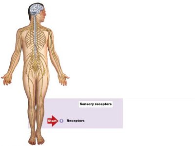
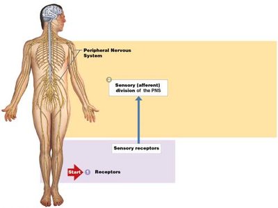
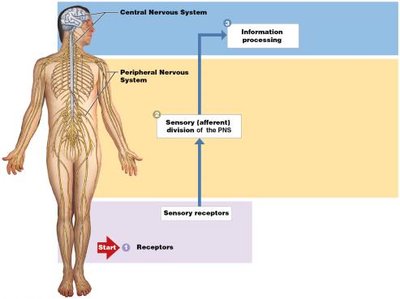
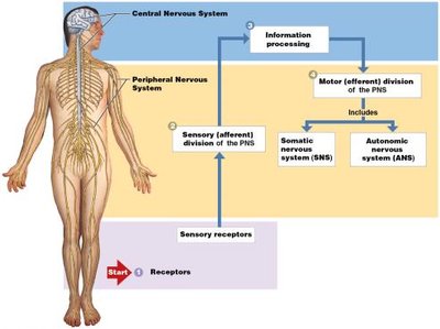
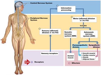
Subdivisions of the Peripheral Nervous System
Sensory (Afferent) Division: Transmits information from receptors in peripheral tissues and organs to the CNS. Includes general sensory receptors (position, touch, pressure, pain, temperature) and special sensory organs (smell, taste, sight, balance, hearing).
Motor (Efferent) Division: Carries motor commands from the CNS to effectors. Subdivided into:
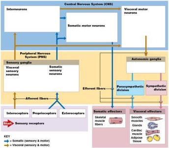
Somatic Nervous System (SNS): Controls voluntary movements of skeletal muscles.
Autonomic Nervous System (ANS): Regulates involuntary activities of smooth muscle, cardiac muscle, glands, and adipose tissue. Further divided into sympathetic and parasympathetic divisions.
General Functions of the Nervous System
Receptors detect changes in the internal or external environment.
Information is sent to the CNS by the sensory division of the PNS.
Information processing (integration and distribution) occurs in the CNS.
Motor commands are carried by the motor division of the PNS.
Effectors respond to commands and alter their activities accordingly.
Neurons: Structure and Function
Structure of a Typical Neuron
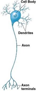
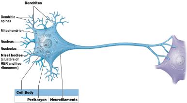
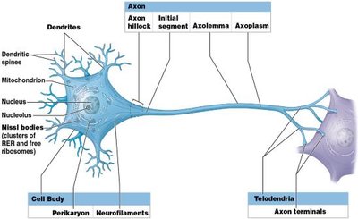
Neurons are specialized cells for intercellular communication, consisting of three main regions:
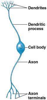
Dendrites: Highly branched processes that receive stimuli from the environment or other neurons.
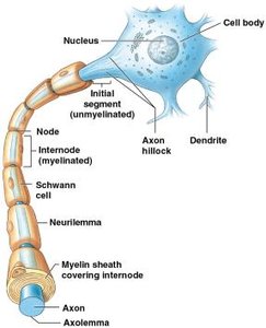
Cell Body (Soma): Contains the nucleus and organelles; responsible for metabolic activities and synthesis of neurotransmitters.
Axon: Conducts electrical impulses (action potentials) toward other cells; originates at the axon hillock and ends in axon terminals (telodendria).
Axoplasmic Transport
Materials such as organelles and neurotransmitters are transported along the axon via neurotubules in a process called axoplasmic transport. This can occur in both directions:
Anterograde transport: From cell body to axon terminals.
Retrograde transport: From axon terminals back to the cell body.
Synapses
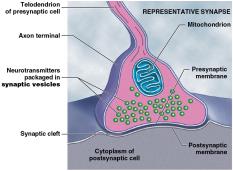
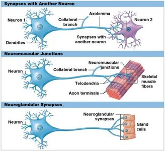
A synapse is the site where a neuron communicates with another cell. The most common type is the chemical synapse, where neurotransmitters are released from the presynaptic membrane into the synaptic cleft and bind to receptors on the postsynaptic membrane.
Neuron Replacement
Most CNS neurons lack centrioles and cannot divide, so they are seldom replaced if lost to injury or disease.
Some neural stem cells exist in the olfactory epithelium, retina, and hippocampus, but most are inactive.
Classification of Neurons
Structural Classification
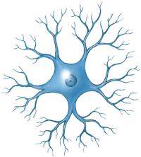
Anaxonic Neurons: Small, indistinguishable axons and dendrites; found in the brain and special sense organs.
Bipolar Neurons: One dendritic process and one axon; rare, found in special sense organs.
Unipolar Neurons: Dendrites and axon are continuous; cell body off to one side; most sensory neurons in the PNS.
Multipolar Neurons: Two or more dendrites and a single axon; most common in the CNS and all motor neurons to skeletal muscles.
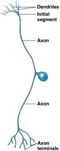
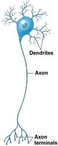
Functional Classification
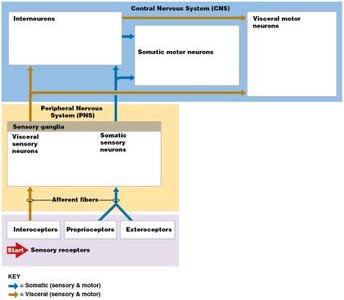
Sensory Neurons: Carry sensory information to the CNS; mostly unipolar; cell bodies in sensory ganglia.
Interneurons: Located in the CNS; connect sensory and motor neurons; involved in higher functions such as memory and learning.
Motor Neurons: Carry instructions from the CNS to effectors; somatic motor neurons control skeletal muscles, visceral motor neurons control smooth/cardiac muscle, glands, and adipose tissue.
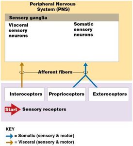
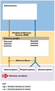
Classification of Sensory Receptors
Interoceptors: Monitor internal organs and systems; detect stretch, deep pressure, and pain.
Proprioceptors: Monitor position and movement of skeletal muscles and joints.
Exteroceptors: Monitor the external environment; detect touch, temperature, pressure, and special senses.
Neuroglia: Support Cells of the Nervous System
Neuroglia of the CNS
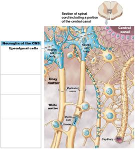
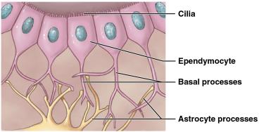
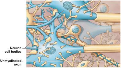
Ependymal Cells: Line the central canal and ventricles; produce, circulate, and monitor cerebrospinal fluid (CSF).
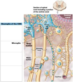
Microglia: Mobile phagocytic cells; remove debris, waste, and pathogens.
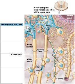
Astrocytes: Maintain the blood-brain barrier, provide structural support, regulate ion/nutrient/gas concentrations, absorb/recycle neurotransmitters, and form scar tissue after injury.
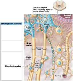
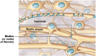
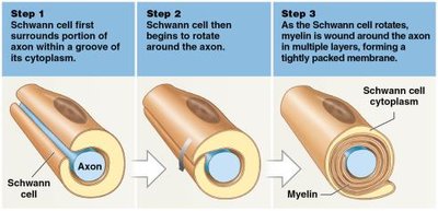
Oligodendrocytes: Stabilize axons and produce myelin sheaths, which speed up nerve impulse transmission.
Neuroglia of the PNS
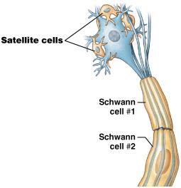
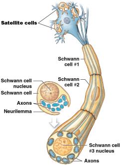
Schwann Cells: Cover peripheral axons; form myelin sheaths and participate in axon repair.
Satellite Cells: Surround peripheral cell bodies; regulate the environment around neurons.
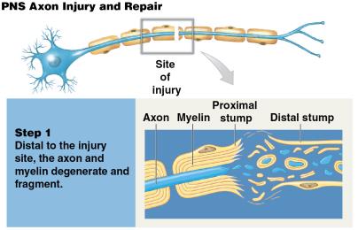
Axon Injury and Repair in the PNS
Axon and myelin degenerate distal to injury (Wallerian degeneration).
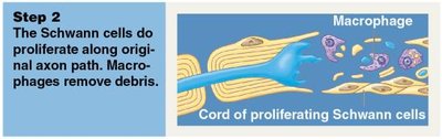
Schwann cells proliferate along the original axon path; macrophages remove debris.
Axon grows along the path created by Schwann cells.
Schwann cells wrap around the elongating axon; normal function may return if synaptic contacts are reestablished.
Neurophysiology: Membrane Potentials and Neural Activity
Membrane Potential and Its Role in Neuronal Activity
Membrane potential is the potential difference across a cell membrane due to unequal charge distribution. The inside of the membrane is slightly negative compared to the outside, primarily due to differences in ion permeability and active transport mechanisms.
Types of Neural Activity
Resting Membrane Potential: The membrane potential of an undisturbed cell (typically –70 mV in neurons).
Graded Potential: Temporary, localized change in resting potential caused by a stimulus; decreases with distance from the stimulus.
Action Potential: Electrical event that starts at one location on the axon membrane and spreads along the axon; triggered by a sufficiently large graded potential.
Synaptic Activity: Neurotransmitter release from the presynaptic cell alters the postsynaptic cell's membrane potential, producing graded potentials.
Resting Membrane Potential: Creation and Maintenance
ECF has high concentrations of Na+ and Cl–; cytosol has high K+ and negatively charged proteins (Pr–).
Passive forces: Diffusion of Na+ and K+ through leak channels; negatively charged proteins cannot diffuse out.
Active process: Sodium–potassium exchange pump ejects 3 Na+ for every 2 K+ recovered, maintaining the resting potential.
Gated Ion Channels and Membrane Permeability
Chemically Gated Channels: Open when bound to specific chemicals (e.g., acetylcholine); abundant on dendrites and cell body.
Voltage-Gated Channels: Open or close in response to changes in membrane potential; found along axons and axon terminals.
Mechanically Gated Channels: Open in response to physical distortion; important in sensory receptors for touch and hearing.
Graded Potentials
Localized changes in membrane potential that cannot spread far from the site of stimulation.
Depolarization: Opening of chemically gated Na+ channels causes the membrane potential to become less negative.
Repolarization: Membrane returns to resting potential as excess Na+ is transported out.
Hyperpolarization: Opening of K+ channels causes the membrane potential to become more negative than resting potential.
Action Potentials
All-or-none events that propagate along the axon.
Depend on voltage-gated ion channels.
Steps:
Depolarization to threshold (about –60 mV).
Activation of sodium channels and rapid depolarization.
Inactivation of sodium channels and activation of potassium channels (repolarization).
Return to resting potential and closure of potassium channels (brief hyperpolarization).
Absolute refractory period: No response possible.
Relative refractory period: Response possible only to a stronger stimulus.
Propagation of Action Potentials
Continuous Propagation: Step-by-step movement along unmyelinated axons; slower.
Saltatory Propagation: Action potential jumps between nodes of Ranvier in myelinated axons; much faster.
Synapses and Information Processing
Chemical and Electrical Synapses
Chemical Synapses: Most common; neurotransmitter release (e.g., acetylcholine at cholinergic synapses) triggers postsynaptic potentials.
Electrical Synapses: Presynaptic and postsynaptic membranes joined by gap junctions; rare, with no variability in response.
Events at a Cholinergic Synapse
Action potential depolarizes axon terminal.
Voltage-gated Ca2+ channels open; Ca2+ triggers ACh release.
ACh binds to receptors on postsynaptic membrane, opening Na+ channels and causing depolarization.
ACh is broken down by acetylcholinesterase (AChE); effects are temporary.
Postsynaptic Potentials and Summation
Excitatory Postsynaptic Potential (EPSP): Graded depolarization; brings membrane closer to threshold.
Inhibitory Postsynaptic Potential (IPSP): Graded hyperpolarization; moves membrane further from threshold.
Summation: Integration of EPSPs and IPSPs; can be temporal (repeated stimulation at one synapse) or spatial (simultaneous stimulation at multiple synapses).
Information Processing in Neurons
Regulatory neurons can facilitate or inhibit presynaptic activity.
Neurotransmitters may have direct or indirect effects on ion channels.
Information is encoded as the frequency of action potentials; higher frequency indicates stronger or more sustained stimulation.
Additional info: This guide covers the foundational concepts of nervous tissue, including cellular organization, neuron structure and function, neuroglia, membrane potentials, synaptic transmission, and information processing, as outlined in a typical Anatomy & Physiology college course.
