 Back
BackNervous Tissue: Structure, Function, and Physiology
Study Guide - Smart Notes
 Tailored notes based on your materials, expanded with key definitions, examples, and context.
Nervous Tissue
An Introduction to the Nervous System
The nervous system is a complex network responsible for communication, coordination, and control throughout the body. It includes the brain, spinal cord, sensory receptors, and nerves that connect to other organ systems.
Neurons: Specialized cells for intercellular communication.
Neuroglia (glial cells): Support, protect, and maintain the environment for neurons.
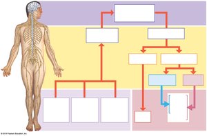
Divisions of the Nervous System
Anatomical Divisions
Central Nervous System (CNS): Brain and spinal cord; processes and coordinates sensory data, motor commands, and higher functions (intelligence, memory, learning, emotion).
Peripheral Nervous System (PNS): All nervous tissue outside the CNS; delivers sensory information to the CNS and carries motor commands to peripheral tissues.
Functional Divisions of the PNS
Afferent Division: Carries sensory information from receptors to the CNS.
Efferent Division: Carries motor commands from the CNS to effectors (muscles, glands, adipose tissue).
Somatic Nervous System (SNS): Controls voluntary and involuntary (reflex) skeletal muscle contractions.
Autonomic Nervous System (ANS): Controls subconscious actions (smooth/cardiac muscle, glands); includes sympathetic (stimulating) and parasympathetic (relaxing) divisions.
Enteric Nervous System (ENS): Network in the digestive tract, capable of local reflexes independent of the CNS but influenced by the ANS.
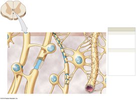
Neurons
Structure and Function
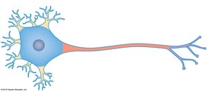
Neurons are the basic functional units of the nervous system, specialized for communication, information processing, and control.
Cell Body (Soma): Contains the nucleus, nucleolus, mitochondria, rough endoplasmic reticulum (RER), and ribosomes.
Dendrites: Short, branched processes that receive signals from other neurons.
Axon: Long process that propagates action potentials to other cells.
Axon Hillock: Region where the axon attaches to the cell body and where action potentials are initiated.
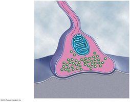
Telodendria: Fine extensions at the end of the axon, ending in axon terminals (synaptic terminals).
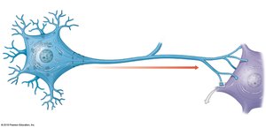
Axonal Transport
Materials move between the cell body and axon terminals via neurotubules, powered by proteins such as kinesin and dynein.
Structural Classification of Neurons
Anaxonic: Small, indistinguishable axons and dendrites; found in brain and sense organs.
Bipolar: One dendrite, one axon; rare, found in special sense organs.
Unipolar: Fused axon and dendrite; most sensory neurons in PNS.
Multipolar: One axon, multiple dendrites; most common in CNS and all motor neurons to skeletal muscle.
Functional Classification of Neurons
Sensory Neurons (Afferent): Transmit sensory information to the CNS.
Motor Neurons (Efferent): Carry instructions from the CNS to effectors.
Interneurons: Connect sensory and motor neurons; involved in integration, memory, and learning.
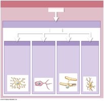
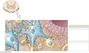
Neuroglia
Types of Neuroglia in the CNS
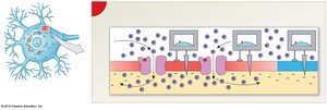
Astrocytes: Maintain the blood-brain barrier, provide structural support, repair tissue, guide development, and regulate the interstitial environment.
Ependymal Cells: Line ventricles and central canal; produce and circulate cerebrospinal fluid (CSF).
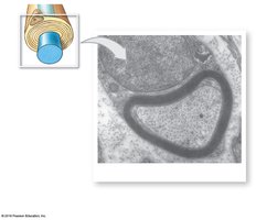
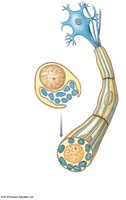
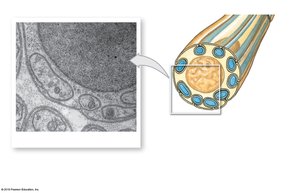
Oligodendrocytes: Myelinate CNS axons, increasing action potential speed; provide structural framework.
Microglia: Phagocytic cells that remove debris, waste, and pathogens.
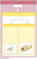
Types of Neuroglia in the PNS
Satellite Cells: Surround neuron cell bodies in ganglia; regulate the environment around neurons.
Schwann Cells: Myelinate peripheral axons; assist in repair after injury.

Neural Responses to Injury
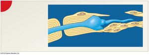
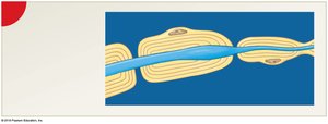
Wallerian Degeneration: Axon distal to injury degenerates; Schwann cells form a path for regrowth.
Regeneration in CNS: Limited by astrocyte activity (scar tissue, inhibitory chemicals).


Membrane Potential
Resting Membrane Potential
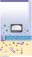
All cells have a membrane potential due to ion movement. In neurons, this is crucial for signal transmission.
Resting Membrane Potential: About –70 mV in neurons; inside is negative relative to outside.
Key Ions: Na+ (outside), K+ (inside), Cl–, and negatively charged proteins (inside).
Sodium-Potassium Pump: Maintains gradients by pumping 3 Na+ out and 2 K+ in, using ATP.
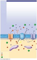
Ion Channels
Passive (Leak) Channels: Always open; permeability varies.
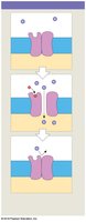
Active (Gated) Channels: Open/close in response to stimuli.
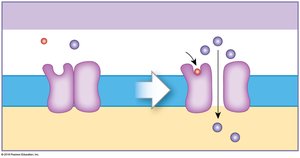
Chemically Gated: Open when specific chemicals bind (e.g., ACh).
Voltage-Gated: Open/close with changes in membrane potential.
Mechanically Gated: Open in response to physical distortion.
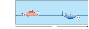
Graded Potentials
Local changes in membrane potential; decrease with distance from stimulus.
Can be depolarizing (Na+ influx) or hyperpolarizing (K+ efflux).
Stronger stimuli produce larger changes.


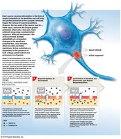
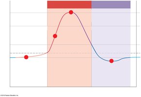
Action Potentials
Generation and Propagation
Action potentials are rapid, propagated changes in membrane potential that travel along axons.
All-or-None Principle: If threshold is reached, an action potential occurs; otherwise, it does not.
Phases:
Depolarization to threshold
Activation of voltage-gated Na+ channels (Na+ influx)
Inactivation of Na+ channels, activation of K+ channels (K+ efflux, repolarization)
Return to resting potential (brief hyperpolarization)
Refractory Periods: Absolute (no new AP possible), Relative (strong stimulus needed).
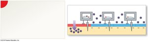
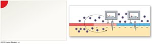
Propagation Types
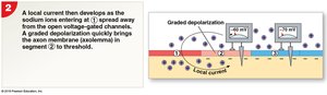
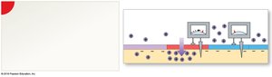
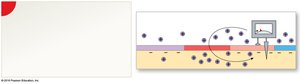
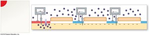
Continuous Propagation: In unmyelinated axons; AP moves segment by segment.
Saltatory Propagation: In myelinated axons; AP jumps from node to node, increasing speed and efficiency.
Axon Types and Conduction Speed
Type A fibers: Large, myelinated, fast (120 m/s); carry critical sensory and motor information.
Type B fibers: Medium, myelinated, intermediate speed (18 m/s).
Type C fibers: Small, unmyelinated, slow (1 m/s); carry less urgent information.
Synapses
Types of Synapses
Electrical Synapses: Direct connection via gap junctions; rapid transmission.
Chemical Synapses: Use neurotransmitters to transmit signals across a synaptic cleft; most common type.
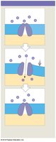
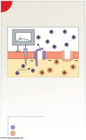
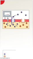
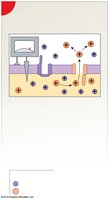
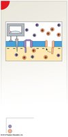
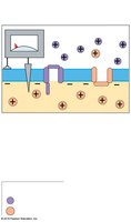
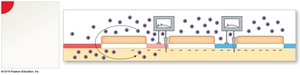
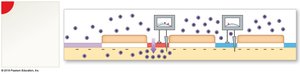
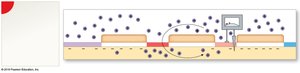
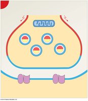
Events at a Cholinergic Synapse
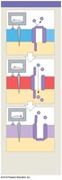
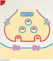
Action potential arrives at axon terminal, depolarizing the membrane.
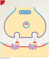
Voltage-gated Ca2+ channels open; Ca2+ triggers exocytosis of ACh.
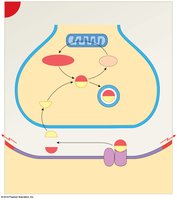
ACh binds to receptors on postsynaptic membrane, causing depolarization.
ACh is broken down by acetylcholinesterase (AChE); choline is reabsorbed and recycled.
Synaptic Delay and Fatigue
Synaptic Delay: Time required for neurotransmitter release and binding (0.2–0.5 ms).
Synaptic Fatigue: Occurs when neurotransmitter supply cannot keep up with demand during intense stimulation.
Neurotransmitters and Neuromodulators
Classes and Effects
Excitatory: Cause depolarization (e.g., glutamate, ACh at neuromuscular junctions).
Inhibitory: Cause hyperpolarization (e.g., GABA).
Biogenic Amines: Norepinephrine, dopamine, serotonin.
Dissolved Gases: Nitric oxide (NO), carbon monoxide (CO).
Neuropeptides: Opioids (endorphins, enkephalins, dynorphins).
Mechanisms of Action
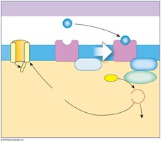
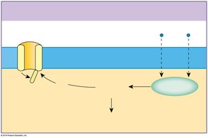
Direct: Open/close ion channels directly (e.g., ACh, glutamate).
Indirect via G Proteins: Activate second messengers (e.g., cAMP) to alter cell activity (e.g., norepinephrine, dopamine).
Indirect via Intracellular Enzymes: Lipid-soluble gases diffuse and activate enzymes inside the cell (e.g., NO, CO).
Information Processing
Postsynaptic Potentials
Excitatory Postsynaptic Potential (EPSP): Graded depolarization; increases likelihood of action potential.
Inhibitory Postsynaptic Potential (IPSP): Graded hyperpolarization; decreases likelihood of action potential.
Summation: EPSPs and IPSPs combine temporally (rapid, repeated stimuli at one synapse) or spatially (simultaneous stimuli at multiple synapses) to determine if threshold is reached.
Presynaptic Modulation
Presynaptic Inhibition: Reduces neurotransmitter release (e.g., GABA).
Presynaptic Facilitation: Increases neurotransmitter release (e.g., serotonin).
Summary
Neural information is relayed as action potentials.
Neurotransmitters and neuromodulators can excite, inhibit, or modulate postsynaptic responses.
Integration of signals at the postsynaptic neuron determines the final response.
