 Back
BackNervous Tissue: Structure, Function, and Physiology
Study Guide - Smart Notes
 Tailored notes based on your materials, expanded with key definitions, examples, and context.
Nervous Tissue and the Nervous System
Introduction to the Nervous System
The nervous system is essential for feeling, thinking, moving, awareness, coordination of body functions, homeostasis, and responsiveness to changes. The main cell type is the neuron, which communicates via electrical impulses. Neurons transmit nerve impulses along nerve fibers to other neurons or cells outside the nervous system at a synapse, where neurotransmitters act as chemical messengers.
Neurons typically have a cell body, an axon, and dendrites.
Nerves are bundles of nerve fibers (axons).
Synapse: Small space between communicating cells.
Neurotransmitters: Chemicals that convey impulses across synapses.
Organization of the Nervous System
The nervous system is divided into two main groups:
Central Nervous System (CNS): Brain and spinal cord.
Peripheral Nervous System (PNS): Nerves connecting CNS to the rest of the body.
General functions include sensory, integrative, and motor activities. Neuroglia provide nutrients, insulation, and support for neurons. 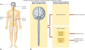
Functional Organization of the Nervous System
General Functions
Sensory function: Sensory receptors gather information and convert it into nerve impulses.
Integrative function: Impulses are processed in the CNS to create sensations, memories, or thoughts.
Motor function: Impulses are sent to effectors (muscles or glands) to perform actions.
Motor functions are divided into:
Somatic nervous system: Voluntary control (skeletal muscles).
Autonomic nervous system: Involuntary control (cardiac & smooth muscle, glands).
Neuron Structure and Classification
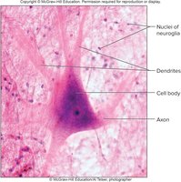
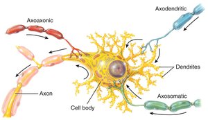
Neuron Structure
Neurons consist of a cell body, dendrites, and an axon.
Dendrites: Conduct impulses toward the cell body; short and branching.
Axon: Conducts impulses away from the cell body; arises from the axon hillock.
Cell body: Contains organelles such as mitochondria, lysosomes, Golgi apparatus, Nissl bodies, neurofilaments, and a nucleus.
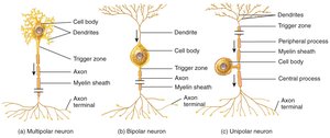
Structural Classification of Neurons
Neurons are classified based on their structure:
Multipolar neurons: Many dendrites, one axon; most common in CNS.
Bipolar neurons: One dendrite, one axon; found in special senses (eyes, nose, ears).
Unipolar neurons: One process splits into two; cell bodies in ganglia outside CNS; sensory neurons.
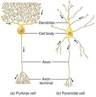
Examples of Dendritic Branching
Dendritic branching varies among neuron types, affecting their connectivity and function.
Purkinje cells: Extensive dendritic trees, found in cerebellum.
Pyramidal cells: Found in cerebral cortex.
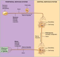
Functional Classification of Neurons
Sensory (afferent) neurons: Conduct impulses from peripheral receptors to CNS; usually unipolar or bipolar.
Motor (efferent) neurons: Conduct impulses from CNS to effectors; multipolar.
Interneurons: Lie within CNS; form links between other neurons; multipolar.
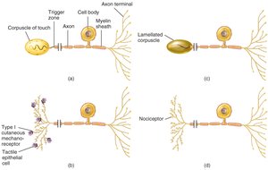
Examples of Sensory Receptors
Sensory receptors are specialized to detect various stimuli, such as touch, pressure, and pain.
Cutaneous mechanoreceptors: Detect touch and pressure.
Nociceptors: Detect pain.
Myelination and Neuroglia
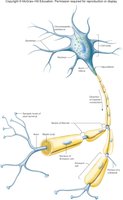
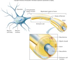
Myelination of Axons
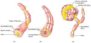
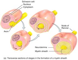
Myelinated fibers: Larger axons are wrapped in myelin sheaths produced by Schwann cells (PNS) or oligodendrocytes (CNS).
Neurilemma: Outer layer of myelin sheath in PNS.
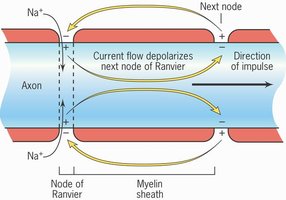
Nodes of Ranvier: Gaps between Schwann cells where ion exchange occurs.
Myelinated and Unmyelinated Axons
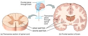
White matter: Regions with many myelinated axons; appears white in CNS.
Gray matter: Regions with neuron cell bodies and unmyelinated axons.
Neuroglia of the CNS
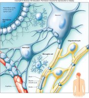
Neuroglia are non-excitable cells that support neurons. Types in CNS:
Microglia: Phagocytize bacteria and debris; produce scar tissue.
Oligodendrocytes: Form myelin sheath in CNS.
Astrocytes: Structural support, regulate nutrients/ions, form blood-brain barrier, scar tissue.
Ependymal cells: Line ventricles and cover choroid plexuses.
Neuroglia of the PNS
Schwann cells: Produce myelin in PNS.
Satellite cells: Structural support and regulation of nutrients/ions.
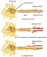
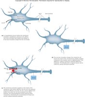
Regeneration and Repair of Neurons
Regeneration in the PNS
Damaged PNS neurons can regenerate if the cell body is intact and Schwann cells are functional. Steps:
Chromatolysis
Wallerian degeneration
Formation of a regeneration tube
Electrical Signals in Neurons
Resting Membrane Potential
Neurons are polarized due to unequal ion distribution.
Inside is more negative than outside.
Resting potential is typically -70 mV.
Maintained by Na+/K+ pump.
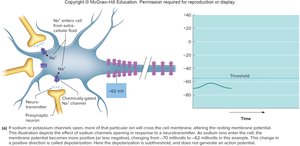
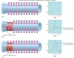
Potential Changes and Action Potentials
Depolarization: Membrane potential becomes less negative.
Threshold potential: -55 mV; triggers action potential.
Repolarization: Return to resting potential.
Hyperpolarization: Membrane becomes overly negative.
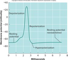
Sequence of Events in an Action Potential
Sodium channels open, Na+ enters cell (depolarization).
Potassium channels open, K+ leaves cell (repolarization).
Resting potential is restored.
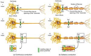
Impulse Conduction
Continuous vs. Saltatory Conduction
Continuous conduction: Unmyelinated fibers; impulse travels along entire membrane.
Saltatory conduction: Myelinated fibers; impulse jumps from node to node, much faster.
Speed is proportional to axon diameter and myelination.
Synapses and Neurotransmitters
The Synapse
Presynaptic neuron: Sends impulse.
Postsynaptic neuron: Receives impulse.
Synaptic knob: Expansion at distal end of presynaptic neuron.
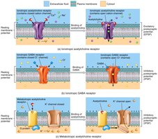
Neurotransmitters released in response to impulse, bind to postsynaptic receptors.
Synaptic Transmission
Neurotransmitters can be excitatory (increase Na+ permeability) or inhibitory (decrease Na+ permeability).
Postsynaptic neuron sums excitatory and inhibitory inputs.
Removal of Neurotransmitter
Diffusion
Enzymatic degradation
Reuptake into cells
Uptake by neuroglial cells
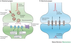
Types of Synapses
Electrical synapse: Gap junctions allow direct transfer of information.
Chemical synapse: One-way transfer via neurotransmitter.
Neurotransmitters and Their Actions
Major Neurotransmitters
Acetylcholine: Excitatory; neuromuscular junctions.
Epinephrine: Excitatory.
GABA, glycine: Inhibitory.
Dopamine, norepinephrine, glutamate, aspartate: Various functions.
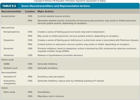
Neurotransmitter | Location | Major Actions |
|---|---|---|
Acetylcholine | CNS/PNS | Stimulates skeletal muscle actions |
Dopamine | CNS | Creates sense of feeling good; deficiency linked to Parkinson's disease |
GABA | CNS | Generally inhibitory |
Glutamate | CNS | Excitatory; important for learning and memory |
Norepinephrine | PNS | Creates sense of well-being; low levels may lead to depression |
Serotonin | CNS | Inhibitory; leads to sleep |
Glycine | CNS | Generally inhibitory |
Endorphins | CNS | Inhibitory; reduce pain |
Impulse Processing and Neural Circuits
Neuronal Pools and Summation
Neurons in the CNS are organized into pools that process information.
Facilitation: Increased neurotransmitter release upon repeated stimulation.
Convergence: Multiple inputs to a single neuron.
Divergence: Single neuron sends impulses to multiple outputs.
Neural Circuits
Reverberating circuit: Later cells repeatedly stimulate early cells (short-term memory).
Parallel after-discharge circuit: Single cell stimulates a group, which all stimulate a common postsynaptic cell (complex processing).
Types of Nerves and Neural Pathways
Types of Nerves
Sensory (afferent) nerves: Bring information to CNS.
Motor (efferent) nerves: Carry impulses from CNS to effectors.
Mixed nerves: Contain both sensory and motor fibers.
Reflex Arcs
Reflex arcs are the simplest neural pathways, providing the basis for involuntary actions. Components:
Sensory receptor
Sensory neuron
Interneuron (reflex center)
Motor neuron
Effector
Regeneration and Plasticity
Regeneration in CNS and PNS
CNS axons rarely regenerate due to inhibitory proteins and scar tissue.
PNS axons can regenerate if conditions are favorable.
Neural Plasticity
Neuroplasticity: Ongoing changes in neurons, glia, and vascular cells.
Synaptic pruning: Elimination of unused or weak connections.
Learning and memory: Short-term memory depends on electrical/chemical events; long-term memory involves structural changes.
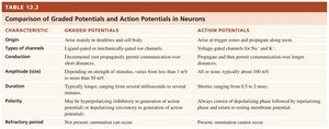
Comparison of Graded and Action Potentials
Characteristic | Graded Potentials | Action Potentials |
|---|---|---|
Origin | Arise mainly in dendrites and cell body | Arise at trigger zones and propagate along axon |
Types of channels | Ligand-gated and mechanically gated | Voltage-gated for Na+ and K+ |
Conduction | Decremental; not propagated | Propagate; communication over longer distances |
Amplitude | Varies with stimulus strength | All-or-none; typically about 100 mV |
Duration | Longer; milliseconds to minutes | Shorter; milliseconds |
Polarity | May be depolarizing or hyperpolarizing | Always consist of depolarizing phase followed by repolarizing phase |
Refractory period | None | Present; summation cannot occur |
Summary
Nervous tissue is fundamental to the function of the nervous system, enabling communication, integration, and response to stimuli. Understanding neuron structure, neuroglia, myelination, synaptic transmission, and neural plasticity is essential for comprehending how the nervous system maintains homeostasis and adapts to changes. Key equations:
Resting membrane potential:
Typical resting potential:
Threshold potential:
Additional info: Academic context was added to clarify structural and functional classifications, synaptic mechanisms, and neural plasticity.
