 Back
BackNervous Tissue: Structure, Function, and Physiology
Study Guide - Smart Notes
 Tailored notes based on your materials, expanded with key definitions, examples, and context.
Introduction to Nervous Tissue
Overview of the Nervous System
The nervous system, together with the endocrine system, regulates and maintains homeostasis in the body. It is responsible for behaviors, memories, and movements. Neurology is the branch of medical science that studies the normal functioning and disorders of the nervous system.
Homeostasis: Maintained by rapid communication and control of body functions.
Functions: Sensing changes, interpreting and remembering those changes, and reacting to them.
Organization of the Nervous System
Major Structures
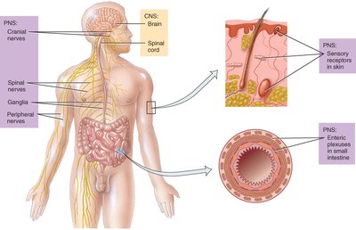
The nervous system is divided into the central nervous system (CNS) and the peripheral nervous system (PNS). The CNS consists of the brain and spinal cord, while the PNS includes cranial nerves, spinal nerves, ganglia, and sensory receptors.
Cranial nerves: 12 pairs emerging from the brain.
Spinal nerves: 31 pairs emerging from the spinal cord.
Ganglia: Clusters of neuron cell bodies outside the CNS.
Sensory receptors: Specialized cells that detect changes in the environment.
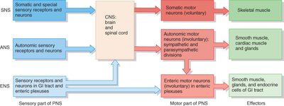
Functional Divisions
Sensory (afferent) function: Detects internal and external stimuli and transmits information to the CNS.
Integrative function: Processes sensory information, stores it, and makes decisions.
Motor (efferent) function: Initiates responses by activating effectors (muscles and glands).
Subdivisions of the PNS
Somatic Nervous System (SNS): Voluntary control of skeletal muscles.
Autonomic Nervous System (ANS): Involuntary control of smooth muscle, cardiac muscle, and glands. Includes sympathetic (fight or flight) and parasympathetic (rest and digest) divisions.
Enteric Nervous System (ENS): Involuntary control of the gastrointestinal tract, functioning independently of the CNS and ANS.
Neuronal Structure and Function
Parts of a Neuron
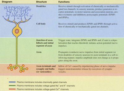
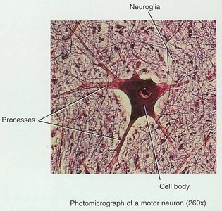
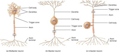
Neurons are the functional units of the nervous system, capable of generating action potentials. They consist of a cell body, dendrites, and an axon.
Cell body (soma): Contains the nucleus and organelles; site of metabolic activity.
Dendrites: Receive incoming signals and conduct impulses toward the cell body.
Axon: Conducts impulses away from the cell body to other neurons or effectors.
Axon hillock: The initial segment where action potentials are generated.
Synaptic end bulbs: Contain neurotransmitters for communication with other cells.
Structural Classification of Neurons
Multipolar: Several dendrites, one axon (most common).
Bipolar: One dendrite, one axon (retina, inner ear, olfactory area).
Unipolar: Single process that splits into two branches (sensory neurons).
Functional Classification of Neurons
Sensory (afferent) neurons: Transmit sensory information to the CNS.
Motor (efferent) neurons: Transmit impulses from the CNS to effectors.
Interneurons (association neurons): Connect sensory and motor neurons within the CNS.
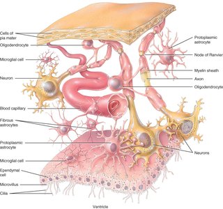
Neuroglial Cells
Types and Functions
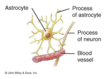
Neuroglia are supporting cells in the nervous system, more numerous than neurons, and capable of cell division. They provide structural and metabolic support, form myelin, and participate in immune defense.
Astrocytes (CNS): Form blood-brain barrier, regulate ion balance, provide structural support.
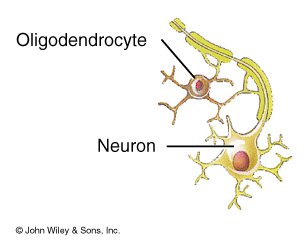
Oligodendrocytes (CNS): Form myelin sheaths around CNS axons.
Microglia (CNS): Phagocytic cells that remove debris and pathogens.
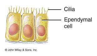
Ependymal cells (CNS): Line ventricles and central canal, produce cerebrospinal fluid (CSF).
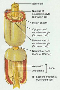
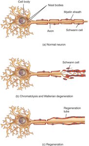
Schwann cells (PNS): Form myelin sheaths around PNS axons.
Satellite cells (PNS): Support neuron cell bodies in ganglia.
Myelination and Axon Coverings
Myelin Sheath
The myelin sheath is a multilayered lipid and protein covering that insulates axons, increasing the speed of nerve impulse conduction. In the PNS, Schwann cells form the myelin sheath, while in the CNS, oligodendrocytes perform this function.
Nodes of Ranvier: Gaps in the myelin sheath where action potentials are regenerated.
Neurolemma: The outermost layer of Schwann cell cytoplasm, important for axon regeneration in the PNS.
Myelinated fibers: Conduct impulses rapidly; appear white.
Unmyelinated fibers: Conduct impulses slowly; only surrounded by neurolemma.
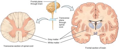
Gray and White Matter
White matter: Composed of myelinated axons; appears white due to myelin.
Gray matter: Contains neuron cell bodies, dendrites, unmyelinated axons, and neuroglia; appears gray.
Distribution: In the spinal cord, gray matter forms an H-shaped core; in the brain, gray matter forms the cortex and nuclei.
Electrical Signals in Neurons
Ion Channels and Membrane Potentials
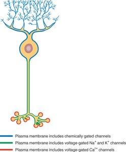
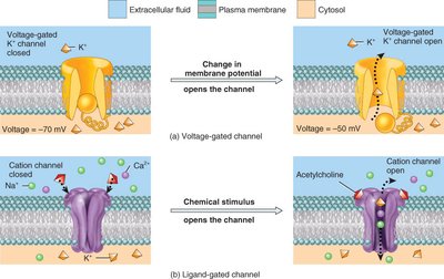
Neurons are electrically excitable due to the presence of ion channels in their membranes. These channels allow the flow of ions, creating electrical signals.
Voltage-gated channels: Open in response to changes in membrane potential.
Ligand-gated channels: Open in response to chemical stimuli (e.g., neurotransmitters).
Mechanically gated channels: Open in response to mechanical deformation (e.g., pressure).
Resting Membrane Potential
The resting membrane potential is the voltage difference across the membrane of a resting neuron, typically -70 mV. It is maintained by differences in ion concentrations inside and outside the cell and selective permeability of the membrane.
Extracellular fluid: High in Na+.
Cytosol: High in K+, organic phosphates, and amino acids.
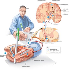
Graded Potentials
Graded potentials are small, localized changes in membrane potential that vary in amplitude and arise mainly in dendrites and cell bodies. They can be depolarizing or hyperpolarizing and are produced by ligand- or mechanically-gated channels.
Action Potentials
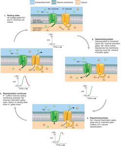
An action potential is a rapid, large change in membrane potential that propagates along the axon. It involves depolarization (Na+ influx) followed by repolarization (K+ efflux). Action potentials follow the all-or-none principle and have refractory periods.
Depolarizing phase: Membrane potential becomes more positive as Na+ enters the cell.
Repolarizing phase: Membrane potential returns to negative as K+ leaves the cell.
After-hyperpolarization: Membrane potential may become more negative than resting potential.
Refractory period: Time during which a neuron cannot generate another action potential (absolute and relative phases).
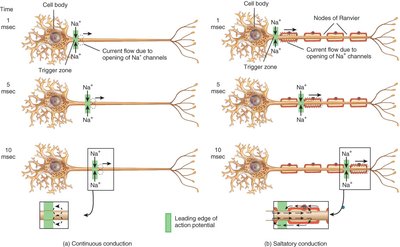
Propagation of Action Potentials
Continuous conduction: Occurs in unmyelinated fibers; action potential moves stepwise along the axon.
Saltatory conduction: Occurs in myelinated fibers; action potential jumps from node to node, increasing speed.
Signal Transmission at Synapses
Types of Synapses
Electrical synapses: Direct passage of ions through gap junctions; fast and bidirectional.
Chemical synapses: Neurotransmitters cross the synaptic cleft to bind receptors on the postsynaptic cell; unidirectional.
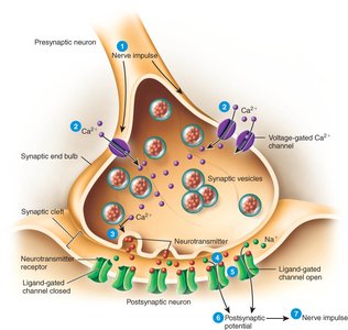
Excitatory and Inhibitory Potentials
Excitatory postsynaptic potential (EPSP): Depolarizes the postsynaptic membrane, increasing the likelihood of an action potential.
Inhibitory postsynaptic potential (IPSP): Hyperpolarizes the postsynaptic membrane, decreasing the likelihood of an action potential.
Removal of Neurotransmitter
Diffusion: Neurotransmitter moves away from the synaptic cleft.
Enzymatic degradation: Breakdown by enzymes (e.g., acetylcholinesterase).
Uptake: Reuptake by neurons or glial cells.
Neurotransmitters
Types and Effects
Acetylcholine (ACh): Excitatory at neuromuscular junctions, inhibitory at others; inactivated by acetylcholinesterase.
Amino acids: Glutamate (excitatory), GABA (inhibitory).
Biogenic amines: Norepinephrine, dopamine, serotonin (regulate mood, sleep, muscle tone).
ATP and purines: Excitatory in CNS and PNS.
Gases: Nitric oxide (NO), acts immediately and diffuses to neighboring cells.
Neuropeptides: Substance P (pain), enkephalins, endorphins (pain relief).
Regeneration and Repair
Plasticity and Repair
Neurons exhibit plasticity, the ability to change and adapt, but have limited capacity for regeneration. In the PNS, axons can regenerate if the cell body is intact and Schwann cells form a regeneration tube. In the CNS, regeneration is very limited.
Disorders of Nervous Tissue
Multiple Sclerosis (MS)
Autoimmune destruction of CNS myelin sheaths, leading to muscle weakness, abnormal sensations, and vision problems.
Characterized by remissions and relapses, with progressive loss of function.
Epilepsy
Characterized by recurrent seizures due to abnormal electrical discharges in the brain.
Causes include brain injury, metabolic disturbances, infections, toxins, and tumors.
