 Back
BackNeural and Chemical Control of Respiration
Study Guide - Smart Notes
 Tailored notes based on your materials, expanded with key definitions, examples, and context.
Control of Respiration
Neural Control of Ventilation
The process of breathing (ventilation) is tightly regulated by neural mechanisms in the brainstem to ensure proper gas exchange and homeostasis. The main centers involved are located in the medulla oblongata and pons, which coordinate the rhythm and depth of breathing.
Dyspnea: The sensation of shortness of breath.
Eupnea: Normal, quiet breathing at rest.
Respiratory Pattern Generator (RPG): A collection of neurons in the superior, anterior medulla that generates the basic rhythm for breathing.
Ventral Respiratory Group (VRG): Located in the anterior and lateral medulla, contains both inspiratory and expiratory neurons. Inspiratory neurons stimulate motor neurons that activate the diaphragm (via the phrenic nerve), external intercostal muscles (via intercostal nerves), and accessory muscles.
Dorsal Respiratory Group (DRG): Found in the posterior medulla, primarily integrates sensory information from the blood and lungs and relays it to other respiratory nuclei. Some DRG neurons are involved in inspiration.
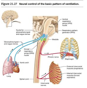
Rate and Depth of Ventilation: Chemoreceptor Control
Ventilation is also regulated by chemoreceptors that monitor the levels of carbon dioxide (PCO2), oxygen (PO2), and hydrogen ions (H+) in the blood and cerebrospinal fluid.
Chemoreceptor: Specialized cell that responds to changes in the concentration of specific chemicals.
Central Chemoreceptors: Located throughout the medulla, midbrain, hypothalamus, and cerebellum. They detect changes in PCO2 and pH by monitoring H+ concentration in the brain's extracellular fluid and cerebrospinal fluid.
Peripheral Chemoreceptors: Found in the carotid bodies (carotid arteries) and aortic bodies (aorta). They detect changes in PCO2, H+, and PO2 in arterial blood.
Arterial PCO2: The most powerful stimulus for changes in ventilation. High PCO2 triggers hyperventilation; low PCO2 triggers hypoventilation.
Example: After intense exercise, increased PCO2 and H+ stimulate hyperventilation to expel excess CO2 and restore homeostasis.
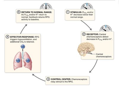
Influence of Peripheral Chemoreceptors
Peripheral chemoreceptors play a crucial role in detecting low oxygen levels (hypoxemia) and contribute to the regulation of ventilation, especially when PO2 falls below 70 mmHg (normal is about 100 mmHg).
Carotid Bodies: Located in the carotid arteries; send signals via the glossopharyngeal nerve (CN IX).
Aortic Bodies: Located in the aorta; send signals via the vagus nerve (CN X).
When PO2 drops, these bodies signal the DRG, which then communicates with the VRG and RPG to increase the rate and depth of ventilation.
Stretch Receptors and Voluntary Control
Additional mechanisms help fine-tune breathing and protect the lungs from damage.
Pulmonary Stretch Receptors: Located in the walls of the trachea and bronchi, these receptors are activated during deep inhalation. They send signals via CN IX and CN X to the DRG, which inhibits inspiratory muscles, preventing over-inflation of the lungs and maintaining eupnea.
Voluntary Control: The cerebral cortex can exert minimal voluntary control over breathing, bypassing the brainstem respiratory centers and sending signals directly through the spinal cord to respiratory muscles. This allows for conscious changes in breathing (e.g., holding your breath), but is limited compared to automatic control.
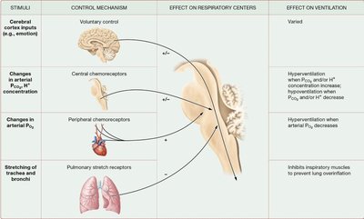
Summary Table: Respiratory Control Mechanisms
Stimuli | Control Mechanism | Effect on Respiratory Centers | Effect on Ventilation |
|---|---|---|---|
Cerebral cortex inputs (e.g., emotion) | Voluntary control | + / - | Varied |
Changes in arterial PCO2, H+ concentration | Central chemoreceptors | + when PCO2 or H+ increases; - when they decrease | Hyperventilation or hypoventilation |
Changes in arterial PO2 | Peripheral chemoreceptors | + when PO2 decreases | Hyperventilation when arterial PO2 decreases |
Stretching of trachea and bronchi | Pulmonary stretch receptors | - | Inhibits inspiratory muscles to prevent lung overinflation |
Key Equations
Relationship between CO2 and pH:
Increased CO2 leads to increased H+ (lower pH, more acidic), stimulating ventilation.
Additional info:
Glossopharyngeal (CN IX) and vagus (CN X) nerves are critical for transmitting sensory information from peripheral chemoreceptors and stretch receptors to the brainstem.
Negative feedback loops are essential for maintaining homeostasis in respiratory control.
