 Back
BackNeural Integration: Sensory and Motor Pathways in the Nervous System
Study Guide - Smart Notes
 Tailored notes based on your materials, expanded with key definitions, examples, and context.
Neural Integration
Terminology and Concepts
Neural integration involves the processing and interpretation of sensory input and the coordination of motor output. Understanding the types and distribution of sensory receptors, as well as the organization of sensory and motor pathways, is essential for comprehending how the nervous system functions.
Receptor: Specialized cell or nerve ending that detects a specific stimulus.
Punctate distribution: Uneven distribution of receptors across the body, resulting in areas of high and low sensitivity.
Adaptation: Reduction in sensitivity to a constant stimulus over time; some receptors adapt quickly, others slowly.
Adaptation of Sensory Receptors
Adaptation refers to the process by which sensory receptors become less responsive to a constant stimulus. This phenomenon allows the nervous system to ignore unimportant stimuli and focus on changes in the environment.
Fast-adapting (phasic) receptors: Respond quickly to stimuli but stop firing if the stimulus remains constant (e.g., Meissner corpuscles, hair root plexus).
Slow-adapting (tonic) receptors: Continue to respond to a constant stimulus (e.g., nociceptors, Merkel discs).
Example: Sustained pressure on the skin is initially felt but soon ignored; pain receptors (nociceptors) adapt poorly, so pain persists.
Classification of Sensory Receptors
By Location
Exteroceptors: Located in the skin; detect external stimuli such as touch and pressure.
Interoceptors: Found in internal organs; detect sensations like pain and pressure from within the body.
Proprioceptors: Located in muscles, tendons, and joints; provide information about body position and movement.
By Stimulus Type
Nociceptors: Detect pain from tissue damage, extreme temperatures, or chemicals; slow-adapting.
Thermoreceptors: Detect changes in temperature; fast-adapting, found in skin, muscles, liver, and hypothalamus.
Mechanoreceptors: Respond to mechanical forces such as touch, pressure, and vibration.
Chemoreceptors: Detect chemical changes in the environment (e.g., taste, smell).
Photoreceptors: Detect light (rods and cones in the eyes).
Types of Mechanoreceptors
Tactile receptors: Include hair root plexus, Merkel discs, Meissner corpuscles, and Pacinian corpuscles; detect touch, pressure, and vibration.
Baroreceptors: Monitor pressure changes in organs and blood vessels; rapidly adapting.
Proprioceptors: Muscle spindles (detect stretch), Golgi tendon organs (detect tension), joint capsule receptors (detect movement and pressure), and internal ear receptors (detect head position).
Referred Pain
Concept and Examples
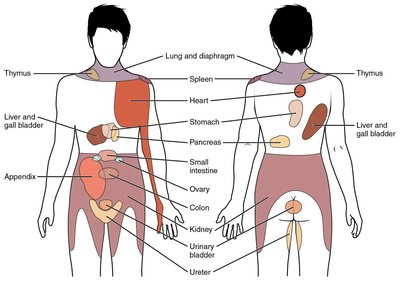
Referred pain occurs when pain is perceived at a location other than the site of the painful stimulus. This phenomenon is due to the convergence of sensory nerve fibers in the spinal cord, leading to misinterpretation by the brain.
Example: Pain from a heart attack may be felt in the left arm; a herniated disk may cause leg pain.
Sensory Pathways
Overview of Sensory Tracts
Sensory information from receptors travels to the brain via specific neural pathways, each responsible for different types of sensations.
Spinothalamic tracts: Carry pain, temperature, crude touch, and pressure sensations.
Posterior columns: Carry discriminative touch, vibration, and proprioception.
Spinocerebellar tracts: Carry proprioceptive information to the cerebellum.
Spinothalamic Tracts
Anterior Spinothalamic Tract: Transmits crude touch and pressure.
Lateral Spinothalamic Tract: Transmits pain and temperature.
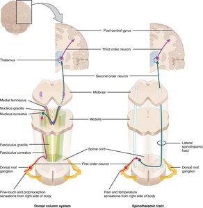
Pathway:
1st neuron: Afferent neuron, cell body in dorsal root ganglion (DRG), synapses in posterior gray horn.
2nd neuron: Cell body in posterior gray horn, crosses to opposite side, ascends to thalamus.
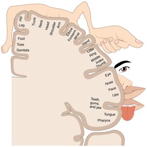
3rd neuron: Cell body in thalamus, projects to postcentral gyrus (primary somatosensory cortex).
Posterior Column Pathway
Function: Transmits discriminative touch, vibration, and proprioception.
Pathway:
1st neuron: Afferent neuron, cell body in DRG, ascends in posterior column to medulla oblongata (no synapse in spinal cord).
2nd neuron: Cell body in medulla, crosses to opposite side, ascends to thalamus.
3rd neuron: Cell body in thalamus, projects to postcentral gyrus.
Spinocerebellar Tracts
Function: Carry proprioceptive information from muscle spindles, Golgi tendon organs, and joint capsules to the cerebellum for coordination of movement.
Motor Pathways
Direct (Pyramidal) Motor Pathways
Direct motor pathways, also known as pyramidal tracts, are responsible for voluntary control of skeletal muscles. They include the corticospinal tracts.
Lateral corticospinal tract: 80% of axons cross at the medullary pyramids and descend in the lateral column.
Anterior corticospinal tract: 20% of axons descend uncrossed and cross at the spinal cord level.
Pathway:
Upper motor neuron (UMN): Cell body in precentral gyrus, axon descends to anterior gray horn.
Lower motor neuron (LMN): Cell body in anterior gray horn, axon exits to innervate skeletal muscle.
Indirect (Extrapyramidal) Motor Pathways
Indirect motor pathways originate outside the precentral gyrus and modulate voluntary movements, muscle tone, and posture. They involve the basal nuclei and reticular formation.
Function: Influence the action of upper motor neurons, initiate or inhibit movements, and regulate muscle tone.
Pathway: Begin in basal nuclei or reticular formation, project to anterior gray horn, and influence lower motor neurons.
Summary Table: Sensory Receptor Types and Functions
Receptor Type | Stimulus Detected | Location | Adaptation |
|---|---|---|---|
Nociceptors | Pain (damage, extreme temp, chemicals) | Skin, organs | Slow |
Thermoreceptors | Temperature | Skin, muscles, liver, hypothalamus | Fast |
Mechanoreceptors | Touch, pressure, vibration, stretch | Skin, muscles, tendons, joints | Varies |
Chemoreceptors | Chemical concentration | Taste buds, olfactory epithelium | Varies |
Photoreceptors | Light | Retina (eye) | Varies |
