 Back
Neural Tissue: Action Potentials, Synaptic Transmission, and Information Processing
Study Guide - Smart Notes
 Tailored notes based on your materials, expanded with key definitions, examples, and context.
Neural Tissue
Action Potentials
Action potentials are rapid, propagated changes in the transmembrane potential that affect the entire excitable membrane of a neuron. They serve as the primary means of communication within the nervous system, linking graded potentials at the cell body with actions at the motor end plate.
Definition: An action potential is a brief reversal of membrane potential, moving from a negative resting value to a positive value and back.
Propagation: Action potentials travel along the axon, ensuring signals reach distant targets efficiently.
All-or-None Principle: An action potential either occurs fully or not at all, depending on whether the threshold is reached.
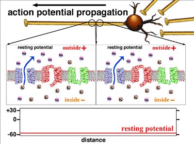
Resting Membrane Potential
The resting membrane potential is the electrical charge difference across the neuron's plasma membrane when the cell is not transmitting a signal. It is typically around -70 mV in neurons.
Key Ions: Sodium (Na+) and potassium (K+) are the main ions involved.
Ion Channels: Voltage-gated sodium and potassium channels are closed at rest.
Distribution: The outside of the membrane is positive relative to the inside, which is negative.
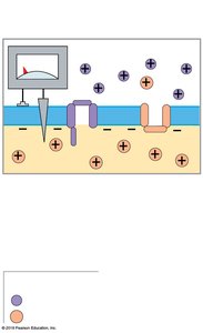
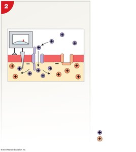
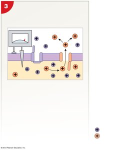
Generation of an Action Potential
The generation of an action potential involves several distinct phases, each characterized by specific ion movements and changes in membrane potential.
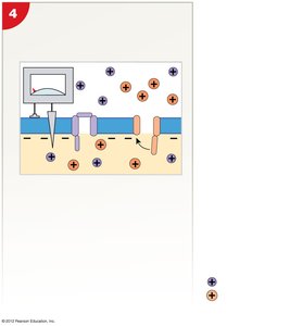
Depolarization to Threshold: A graded depolarization at the axon hillock brings the membrane potential to the threshold (about -60 to -55 mV), triggering the opening of voltage-gated Na+ channels.
Rapid Depolarization: Na+ ions rush into the cell, causing the inner membrane to become positive (up to +30 mV).
Repolarization: At +30 mV, Na+ channels inactivate and K+ channels open, allowing K+ to exit and the membrane potential to return toward negative values.
Hyperpolarization: K+ channels may remain open slightly longer, causing the membrane potential to become more negative than the resting level (down to -90 mV) before returning to rest.
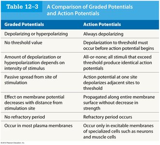
Comparison of Graded Potentials and Action Potentials
Graded potentials and action potentials are two types of changes in membrane potential, each with distinct properties and roles in neural signaling.
Graded Potentials | Action Potentials |
|---|---|
Depolarizing or hyperpolarizing | Always depolarizing |
No threshold value | Depolarization to threshold must occur before action potential begins |
Amplitude depends on stimulus intensity | All-or-none; identical action potentials if threshold exceeded |
Passive spread from site of stimulation | Propagated along entire membrane without decrease in strength |
Effect decreases with distance | No decrease with distance |
No refractory period | Refractory period occurs |
Occur in most plasma membranes | Occur only in excitable membranes (neurons, muscle cells) |
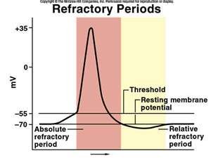
Refractory Periods
The refractory period is the time during which a neuron is unable or less able to fire another action potential. It ensures unidirectional propagation and limits the frequency of action potentials.
Absolute Refractory Period: No action potential can be generated, regardless of stimulus strength (Na+ channels open or inactivated).
Relative Refractory Period: A stronger-than-normal stimulus can initiate another action potential (membrane potential almost normal, some K+ channels still open).
All-or-None Principle
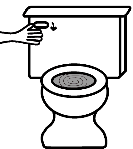
The all-or-none principle states that once the threshold is reached, an action potential will always occur with the same amplitude and duration, regardless of the strength of the stimulus above threshold. This is analogous to flushing a toilet: once the handle is pressed past a certain point, the flush occurs completely.
Propagation of Action Potentials
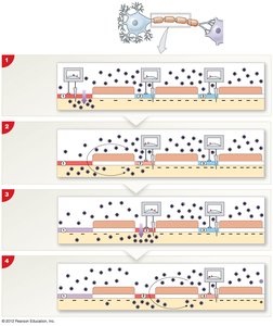
Action potentials propagate along axons in two main ways: continuous propagation (unmyelinated axons) and saltatory propagation (myelinated axons).
Continuous Propagation: Action potential moves along every segment of the membrane, slower due to sequential opening of channels.
Saltatory Propagation: Action potential jumps from node to node (Nodes of Ranvier) in myelinated axons, greatly increasing speed.
Axon Diameter and Speed
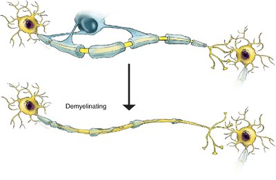
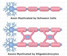
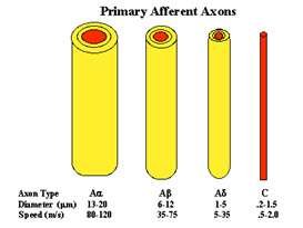
The speed of action potential propagation depends on axon diameter and myelination. Larger diameter and myelinated axons conduct impulses faster due to lower resistance and saltatory conduction.
Type A fibers: Large, myelinated, fastest (e.g., motor neurons).
Type B fibers: Medium, myelinated, intermediate speed.
Type C fibers: Small, unmyelinated, slowest (e.g., pain fibers).
Synapses
Types of Synapses
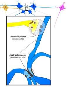
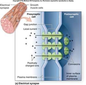
Synapses are specialized junctions where neurons communicate with other neurons or effector cells. There are two main types:
Electrical Synapses: Direct physical contact via gap junctions, allowing ions to pass directly between cells. Rare, but found in some brain regions and the eye.
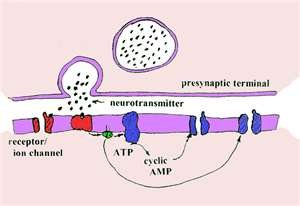
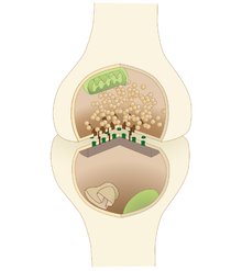
Chemical Synapses: Most common; use neurotransmitters to transmit signals across a synaptic cleft. Action potential in presynaptic neuron triggers neurotransmitter release, which binds to receptors on the postsynaptic cell.
Neurotransmitters and Their Effects
Neurotransmitters are chemicals released by neurons to transmit signals across synapses. Their effect depends on the receptor type, not the neurotransmitter itself.
Excitatory Neurotransmitters: Cause depolarization and promote action potentials (e.g., glutamate, acetylcholine).
Inhibitory Neurotransmitters: Cause hyperpolarization and suppress action potentials (e.g., GABA).

Cholinergic Synapses
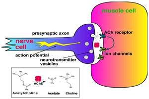
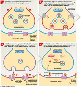
Cholinergic synapses release acetylcholine (ACh) and are found at neuromuscular junctions, many CNS synapses, and all neuron-to-neuron synapses in the PNS and parasympathetic division of the ANS.
Events: Action potential arrives, Ca2+ influx triggers ACh release, ACh binds to receptors, and is broken down by acetylcholinesterase (AChE).
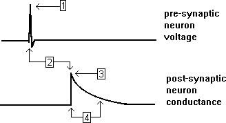
Synaptic Delay: Time between arrival of action potential and postsynaptic effect (0.2–0.5 ms).
Synaptic Fatigue: Occurs when neurotransmitter cannot be recycled fast enough during intense stimulation.
Important Neurotransmitters
Several neurotransmitters play key roles in the nervous system:
Norepinephrine (NE): Excitatory, released by adrenergic synapses, important in CNS and ANS.
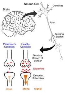
Dopamine (DA): Can be excitatory or inhibitory; involved in movement control and reward pathways. Deficiency linked to Parkinson's disease.
Serotonin: Affects mood, attention, and emotional states; targeted by SSRIs (e.g., Prozac).
GABA: Main inhibitory neurotransmitter in the CNS; reduces anxiety.
Gaseous Neurotransmitters: Nitric oxide (NO) and carbon monoxide (CO) act as neuromodulators in specific tissues.
Neuromodulators
Neuromodulators are chemicals released by synaptic terminals that modulate synaptic transmission. They often have long-term, slow effects and can influence both presynaptic and postsynaptic membranes.
Neuropeptides: Small peptides that activate enzymes via receptor binding.
Opioids: CNS neuromodulators that bind to opiate receptors and relieve pain (e.g., endorphins).
Mechanisms of Neurotransmitter Function
Neurotransmitters can act via direct or indirect mechanisms:
Ionotropic Effects: Directly alter ion movement through ligand-gated channels (e.g., ACh, glutamate).
Indirect Effects via G Proteins: Activate second messenger systems (e.g., cAMP) to alter cell metabolism (e.g., NE, dopamine).
Indirect Effects via Intracellular Enzymes: Gaseous neurotransmitters (NO, CO) activate intracellular enzymes to produce secondary messengers.
Information Processing in Neural Tissue
Postsynaptic Potentials
Postsynaptic potentials are graded changes in the membrane potential of the postsynaptic cell in response to neurotransmitter binding.
Excitatory Postsynaptic Potential (EPSP): Graded depolarization that brings the membrane closer to threshold.
Inhibitory Postsynaptic Potential (IPSP): Graded hyperpolarization that moves the membrane further from threshold.
Summation
Summation is the process by which multiple postsynaptic potentials combine to influence the likelihood of an action potential.
Temporal Summation: Multiple stimuli from a single source in rapid succession add together.
Spatial Summation: Simultaneous stimuli from multiple sources at different locations combine their effects.
Presynaptic Modulation
Presynaptic inhibition and facilitation occur at axoaxonic synapses, where one neuron modulates the neurotransmitter release of another.
Presynaptic Inhibition: Reduces neurotransmitter release, decreasing postsynaptic effect (e.g., GABA-mediated).
Presynaptic Facilitation: Increases neurotransmitter release, enhancing postsynaptic effect (e.g., serotonin-mediated).
Rate of Action Potential Generation
The frequency of action potentials depends on the degree of depolarization above threshold. The maximum rate is limited by the refractory periods, with large-diameter axons having the shortest absolute refractory periods.
