 Back
BackNeural Tissue and the Nervous System: Structure, Function, and Physiology
Study Guide - Smart Notes
 Tailored notes based on your materials, expanded with key definitions, examples, and context.
Neural Tissue and the Nervous System
Introduction to the Nervous System
The nervous system is responsible for coordinating all body activities by transmitting signals between different parts of the body. It consists of specialized cells called neurons and supporting cells known as neuroglia (or glial cells). The nervous system includes the brain, spinal cord, sensory receptors, and nerves that connect these organs to the rest of the body.
Neurons: Cells that send and receive electrical signals.
Neuroglia: Cells that support, protect, and nourish neurons.
Organs: Brain, spinal cord, sensory receptors (e.g., eyes, ears), and nerves.
General Characteristics of Neural Tissue
Neural tissue is highly specialized for communication and control. Its main characteristics include:
Excitability: Ability to generate electrical impulses (action potentials).
Conductivity: Ability to transmit electrical impulses over distances.
Specialization: Neurons are highly specialized and do not divide in adults.
Divisions of the Nervous System
Anatomical Divisions
The nervous system is divided into two main anatomical divisions:
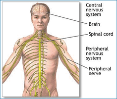
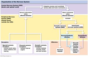
Central Nervous System (CNS): Consists of the brain and spinal cord. It processes, integrates, and coordinates sensory data and motor commands. It is also responsible for higher functions such as intelligence, memory, learning, and emotions.
Peripheral Nervous System (PNS): Includes all neural tissue outside the CNS. It delivers sensory information to the CNS and carries motor commands to peripheral tissues and systems via cranial and spinal nerves.
Functional Divisions of the PNS
The PNS is further divided based on function:
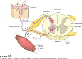
Afferent Division: Carries sensory information from receptors to the CNS.
Efferent Division: Carries motor commands from the CNS to effectors (muscles, glands, adipose tissue).
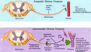
Somatic Nervous System (SNS): Controls voluntary and involuntary (reflex) skeletal muscle contractions.
Autonomic Nervous System (ANS): Controls involuntary actions of smooth muscle, cardiac muscle, glands, and adipose tissue. It is subdivided into the sympathetic (fight-or-flight) and parasympathetic (rest-and-digest) divisions.
Neuron Structure and Classification
Structure of a Neuron
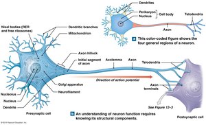
Neurons are the basic functional units of the nervous system. They have specialized structures for receiving, processing, and transmitting information:
Dendrites: Receive information from other neurons.
Cell Body (Soma): Contains the nucleus, nucleolus, mitochondria, and Nissl bodies (rough endoplasmic reticulum and free ribosomes).
Axon Hillock: Region where the axon originates from the cell body.
Axon: Conducts action potentials away from the cell body to other neurons or effectors.
Axon Terminals: Store neurotransmitters in synaptic vesicles for release at synapses.
Structural Classification of Neurons
Bipolar Neurons: One dendrite and one axon; found in special sense organs (rare).
Unipolar Neurons: Fused dendrite and axon with cell body to the side; sensory neurons of the PNS.
Multipolar Neurons: Multiple dendrites and one axon; most common type, found in CNS and as motor neurons.
Functional Classification of Neurons
Sensory (Afferent) Neurons: Monitor internal and external environments and transmit information to the CNS.
Motor (Efferent) Neurons: Carry instructions from the CNS to effectors (muscles, glands, adipose tissue).
Interneurons: Located between sensory and motor neurons; responsible for integration, memory, planning, and learning.
Neuroglia (Glial Cells)
CNS Neuroglia
Neuroglia in the CNS support and protect neurons, and perform various specialized functions:
Ependymal Cells: Line the central canal of the spinal cord and ventricles of the brain; secrete and circulate cerebrospinal fluid (CSF).
Astrocytes: Maintain the blood-brain barrier, repair damaged neural tissue, and regulate the extracellular environment.
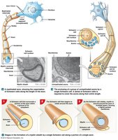
Oligodendrocytes: Form myelin sheaths around CNS axons, increasing the speed of action potentials.
Microglia: Migrate through neural tissue, removing debris, waste, and pathogens by phagocytosis.
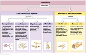
PNS Neuroglia
Satellite Cells: Surround neuron cell bodies in ganglia, regulating the environment around neurons.
Schwann Cells: Form myelin sheaths around PNS axons; can enclose unmyelinated axons as well.
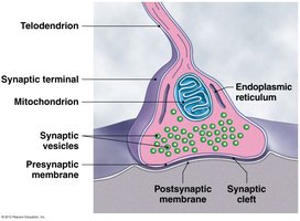
Synapses and Neurotransmission
Synapse Structure and Function
A synapse is the site where a neuron communicates with another cell. The presynaptic cell sends the signal, and the postsynaptic cell receives it. The synaptic cleft separates the two cells.
Presynaptic Terminal: Contains synaptic vesicles filled with neurotransmitters.
Neurotransmitter Release: Triggered by an action potential, neurotransmitters are released into the synaptic cleft and bind to receptors on the postsynaptic membrane.
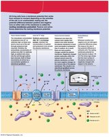
Transmembrane Potential and Action Potentials
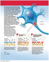
Resting Membrane Potential
Neurons maintain a resting membrane potential due to differences in ion concentrations across the plasma membrane and selective permeability. The typical resting potential is −70 mV.
Extracellular Fluid: High in Na+ and Cl−.
Intracellular Fluid: High in K+ and negatively charged proteins.
Sodium-Potassium Pump: Maintains gradients by pumping 3 Na+ out and 2 K+ in per cycle.
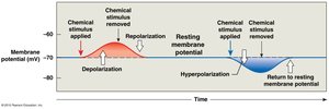
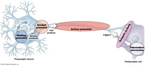
Graded Potentials
Graded potentials are local changes in membrane potential that decrease with distance from the stimulus. They are produced by any stimulus that opens a gated channel.
Depolarization: Opening of Na+ channels causes the membrane potential to become more positive.
Repolarization: Removal of the stimulus returns the membrane potential to resting levels.
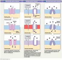
Action Potentials
Action potentials are rapid, propagated changes in membrane potential that travel along the axon. They follow the all-or-none principle: if the threshold is reached, an action potential is generated.
Depolarization to Threshold: Graded depolarization opens voltage-gated Na+ channels.
Rapid Depolarization: Na+ ions rush in, making the inside positive.
Repolarization: Na+ channels close, K+ channels open, K+ exits the cell.
Return to Resting Potential: K+ channels close, membrane returns to −70 mV.
Propagation of Action Potentials
Action potentials propagate along axons in two ways:
Continuous Propagation: Occurs in unmyelinated axons; action potential moves along every part of the membrane.
Saltatory Propagation: Occurs in myelinated axons; action potential jumps from node to node, increasing speed and efficiency.
Axon Diameter: Larger diameter axons conduct impulses faster due to lower resistance.
Neurotransmitters and Synaptic Integration
Types of Neurotransmitters
Excitatory: Cause depolarization and promote action potentials (e.g., acetylcholine at most synapses).
Inhibitory: Cause hyperpolarization and suppress action potentials (e.g., GABA).
Biogenic Amines: Norepinephrine (excitatory), dopamine (excitatory or inhibitory), serotonin (modulates mood and attention).
Dissolved Gases: Nitric oxide, carbon monoxide.
Synaptic Integration and Information Processing
Neurons integrate thousands of excitatory and inhibitory inputs. If the net effect at the axon hillock reaches threshold, an action potential is generated.
Excitatory Postsynaptic Potentials (EPSP): Graded depolarizations caused by opening of chemically-gated Na+ channels.
Inhibitory Postsynaptic Potentials (IPSP): Graded hyperpolarizations caused by opening of chemically-gated K+ channels.
Clinical Applications
Multiple Sclerosis (MS): Progressive demyelination of CNS axons, leading to vision loss, speech, balance, and motor problems.
Guillain-Barré Syndrome: Autoimmune demyelination of PNS axons, causing temporary paralysis.
Diptheria: Bacterial toxins damage Schwann cells, potentially causing fatal paralysis.
