 Back
BackNeuroglia, Myelin Sheath, and Electrophysiology of Neurons: Study Notes
Study Guide - Smart Notes
 Tailored notes based on your materials, expanded with key definitions, examples, and context.
CH 11 PT 2 - Neuroglia of the Central and Peripheral Nervous System
Neuroglia in the CNS
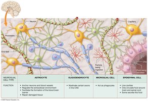
Neuroglia are specialized support cells in the nervous system, essential for maintaining neuronal health and function. The CNS contains several types of neuroglia, each with distinct roles:
Oligodendrocytes: Form concentric layers of plasma membrane called myelin around axons, creating the myelin sheath that increases the speed of nerve impulse conduction.
Microglia: Small, branching cells activated by brain injury; act as wandering phagocytes, ingesting pathogens, dead neurons, and debris, and stimulating inflammation.
Ependymal Cells: Ciliated cells that circulate cerebrospinal fluid (CSF) in the brain and spinal cord; some produce CSF, others monitor its composition.
Astrocytes (not detailed in the provided text but commonly included) anchor neurons and blood vessels, regulate the extracellular environment, and contribute to the blood-brain barrier.
COVID-19 Connection: Microglia and Brain Fog
Post-infection with SARS-CoV-2, about one in four survivors experience "brain fog," characterized by slowed cognitive processing and difficulties with memory, attention, and concentration. This may be due to increased microglial activity, which reduces oligodendrocyte precursors and myelin, slowing impulse conduction.
Neuroglia in the PNS
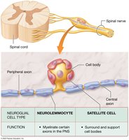
Neurolemmocytes (Schwann Cells): Encase axons in the PNS, forming myelin sheaths and aiding in axon repair.
Satellite Cells: Flat cells surrounding neuron cell bodies in the PNS, supporting and regulating the extracellular environment.
The Myelin Sheath
Structure and Function
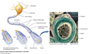
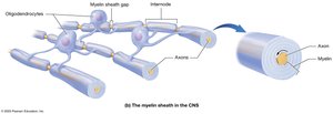
The myelin sheath is formed by neurolemmocytes in the PNS and oligodendrocytes in the CNS. It consists of repeating layers of plasma membrane rich in phospholipids, cholesterol, and proteins, acting as an insulator to prevent ion movement and greatly increase the speed of action potential conduction.
Myelinated axons conduct action potentials 15–150 times faster than unmyelinated axons.
Myelination begins early in the fetal period in the PNS, but much later in the CNS, especially in the brain.
Myelination Process
PNS: Neurolemmocytes wrap outward around a single axon, forming up to 100 layers. The outermost layer is called the neurolemma.
CNS: Oligodendrocyte processes wrap inward, and one cell can myelinate multiple axons.
Segments covered by myelin are called internodes; gaps between them are nodes of Ranvier.
Unmyelinated Axons and White/Gray Matter
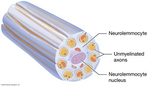
Short axons are typically unmyelinated.
In the PNS, unmyelinated axons are still associated with neurolemmocytes.
In the CNS, myelinated areas appear as white matter, while unmyelinated areas are gray matter.
Regeneration of Nervous Tissue
Regeneration in the CNS and PNS
Regeneration is the replacement of damaged tissue with original tissue. In humans, nervous tissue regeneration is limited:
CNS: Dendrites and axons rarely regenerate due to inhibitory oligodendrocytes, absence of growth factors, and astrocyte-induced scar tissue.
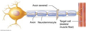
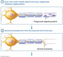
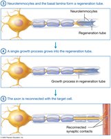
PNS: Regeneration is possible if the cell body remains intact and conditions are ideal. The process involves:
Degeneration of the axon and myelin sheath distal to injury (Wallerian Degeneration).
Phagocytes digest debris.
Growth processes form from the proximal end of the axon.
Neurolemmocytes and basal lamina form a regeneration tube.
A single growth process grows into the tube, reconnecting the axon with the target cell.
Electrophysiology of Neurons
Excitability and Conductivity
Neurons are excitable cells that respond to stimuli (chemical, electrical, mechanical) by generating electrical changes across their plasma membranes. These changes are rapidly conducted along the membrane.
Local Potentials: Short-distance electrical changes.
Action Potentials: Long-distance signals traveling the entire axon.
Resting Membrane Potential
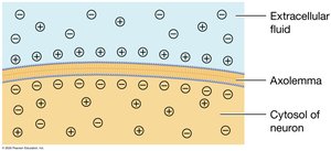
The membrane potential is the electrical gradient across the plasma membrane. At rest, neurons have a resting membrane potential of about -70 mV, due to the loss of potassium ions through leak channels. The cell is polarized at rest.
Ion Channels and Gradients
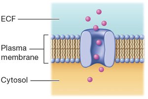
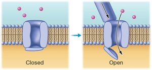
Ions cross the plasma membrane via protein channels or pumps:
Leak Channels: Always open, allow passive ion movement.
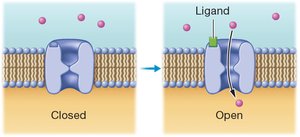
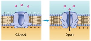
Gated Channels: Open/close in response to specific stimuli:
Ligand-Gated: Open when a chemical (ligand) binds.
Voltage-Gated: Open/close with changes in membrane potential.
Mechanically Gated: Open/close with mechanical stimulation.
Sodium-Potassium Pump
The sodium-potassium pump maintains ion gradients by moving two potassium ions into the cytosol and three sodium ions out to the extracellular fluid, consuming ATP.
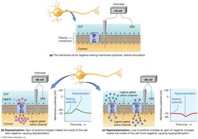
Changes in Membrane Potential
Depolarization: Influx of cations (e.g., sodium) makes the membrane potential less negative.
Repolarization: Return to resting membrane potential.
Hyperpolarization: Membrane potential becomes more negative than at rest.
Local Potentials
Local (graded) potentials are small, reversible changes in membrane potential. They are decremental, meaning the current dissipates over a short distance.
Action Potentials
An action potential is a rapid, uniform depolarization and repolarization of the membrane potential, generated in the axon's trigger zone. Dendrites and cell bodies only generate local potentials.
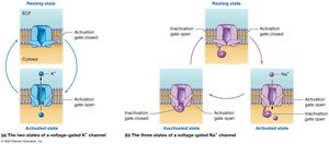
States of Voltage-Gated Channels
Potassium Channels: Resting (closed) and activated (open) states.
Sodium Channels: Two gates (activation and inactivation) and three states: resting, activated, and inactivated.
Summary Table: Types of Ion Channels
Channel Type | Stimulus | Function |
|---|---|---|
Leak Channel | None (always open) | Passive ion movement |
Ligand-Gated Channel | Chemical (ligand) binding | Allows ions to flow in response to neurotransmitters |
Voltage-Gated Channel | Change in membrane potential | Initiates action potentials |
Mechanically Gated Channel | Mechanical stimulation | Responds to touch, pressure, vibration |
Summary Table: Neuroglial Cell Types and Functions
Cell Type | Location | Function |
|---|---|---|
Oligodendrocyte | CNS | Myelinate axons |
Microglia | CNS | Phagocytosis, immune response |
Ependymal Cell | CNS | Circulate and produce CSF |
Neurolemmocyte (Schwann Cell) | PNS | Myelinate axons, aid in repair |
Satellite Cell | PNS | Support cell bodies, regulate environment |
Additional info: Astrocytes, though not detailed in the provided text, are a major neuroglial cell type in the CNS, involved in support, regulation, and repair.
