 Back
BackNeurons: Cellular and Network Properties & The Central Nervous System
Study Guide - Smart Notes
 Tailored notes based on your materials, expanded with key definitions, examples, and context.
Neurons: Cellular and Network Properties
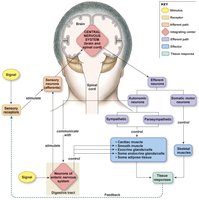
Organization of the Nervous System
The nervous system is divided into the central nervous system (CNS) and the peripheral nervous system (PNS). The CNS consists of the brain and spinal cord, while the PNS includes all neural tissue outside the CNS. The nervous system is responsible for receiving sensory input, integrating information, and coordinating motor output.
Afferent (sensory) neurons carry information to the CNS.
Efferent (motor) neurons carry commands from the CNS to effectors such as muscles and glands.
The efferent division is further divided into the somatic motor (voluntary control of skeletal muscles) and autonomic (involuntary control of smooth muscle, cardiac muscle, and glands) branches.
The autonomic division includes the sympathetic and parasympathetic branches.
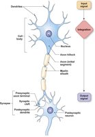
Structure of a Model Neuron
Neurons are the functional units of the nervous system. They consist of a cell body (soma), dendrites, and an axon. Dendrites receive incoming signals, while the axon transmits outgoing electrical impulses to other cells.
Dendrites: Receive input from other neurons.
Axon: Conducts action potentials away from the cell body.
Axon hillock: Site where action potentials are initiated.
Myelin sheath: Insulates axons to speed up signal transmission.
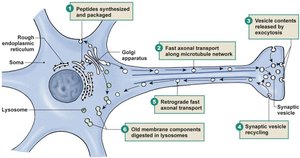
Axonal Transport
Axons transport materials between the cell body and axon terminals using two main mechanisms:
Fast axonal transport: Moves organelles and vesicles rapidly along microtubules (up to 400 mm/day).
Slow axonal transport: Moves cytosolic and cytoskeletal proteins more slowly (0.2–2.5 mm/day).
Transport can be anterograde (from cell body to axon terminal) or retrograde (from axon terminal to cell body).
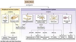
Glial Cells and Their Functions
Glial cells support neurons structurally and functionally. They are found in both the CNS and PNS and have various roles, including forming myelin, providing nutrients, and maintaining homeostasis.
Schwann cells (PNS): Form myelin sheaths around peripheral axons.
Oligodendrocytes (CNS): Form myelin sheaths around multiple CNS axons.
Astrocytes (CNS): Maintain the blood-brain barrier, provide nutrients, and regulate the extracellular environment.
Microglia (CNS): Act as immune cells.
Ependymal cells (CNS): Line ventricles and produce cerebrospinal fluid.
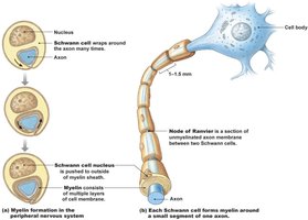
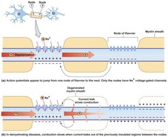
Myelination in the Nervous System
Myelin sheaths are formed by Schwann cells in the PNS and oligodendrocytes in the CNS. Myelination increases the speed of action potential conduction along axons by insulating them and allowing saltatory conduction between nodes of Ranvier.
Each Schwann cell wraps around a single axon segment in the PNS.
Oligodendrocytes can myelinate multiple axons in the CNS.
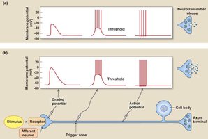
Electrical Signals in Neurons: Graded Potentials vs. Action Potentials
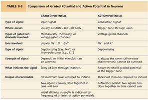
Neurons communicate via electrical signals: graded potentials and action potentials. Graded potentials are variable-strength signals that travel short distances, while action potentials are all-or-none signals that travel long distances along axons.
Graded Potential | Action Potential | |
|---|---|---|
Type of signal | Input signal | Conduction signal |
Where occurs | Dendrites and cell body | Trigger zone through axon |
Types of gated ion channels | Mechanically, chemically, or voltage-gated | Voltage-gated |
Ions involved | Na+, K+, Ca2+, Cl- | Na+, K+ |
Type of signal | Depolarizing or hyperpolarizing | Depolarizing |
Strength of signal | Varies; can be summed | Always the same; all-or-none |
What initiates the signal | Entry of ions through channels | Above-threshold graded potential at trigger zone |
Unique characteristics | No minimum level required; signals can sum | Refractory period prevents summation |
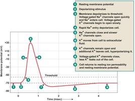
Action Potential: Phases and Ion Movements
An action potential is a rapid, temporary change in membrane potential that propagates along the axon. It involves the sequential opening and closing of voltage-gated Na+ and K+ channels.
Depolarization: Na+ channels open, Na+ enters the cell.
Repolarization: K+ channels open, K+ leaves the cell.
Hyperpolarization: K+ channels remain open, membrane potential becomes more negative than resting.
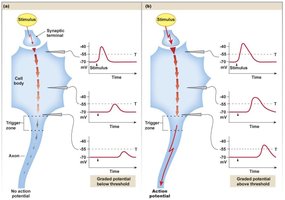
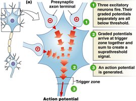
Graded Potentials and Action Potentials: Relationship
Graded potentials occur in the dendrites and cell body and can summate to trigger an action potential if the threshold is reached at the axon hillock (trigger zone). Action potentials are then propagated along the axon.
Coding for Stimulus Intensity
The intensity of a stimulus is encoded by the frequency of action potentials. A stronger stimulus generates a higher frequency of action potentials, not a larger amplitude.
Myelinated Axons and Saltatory Conduction
Myelinated axons conduct action potentials more rapidly due to saltatory conduction, where the action potential jumps from one node of Ranvier to the next. Demyelinating diseases, such as multiple sclerosis, slow or block conduction.
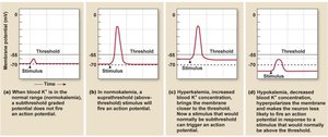
Chemical Factors Affecting Neuronal Excitability
Extracellular potassium concentration significantly affects the excitability of neurons. Hyperkalemia (high K+) increases excitability, while hypokalemia (low K+) decreases it.
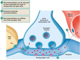
Chemical Synapses and Neurotransmitter Release
Most neuron-to-neuron communication occurs at chemical synapses, where neurotransmitters are released from the presynaptic neuron and bind to receptors on the postsynaptic cell. Calcium influx into the axon terminal triggers exocytosis of neurotransmitter vesicles.
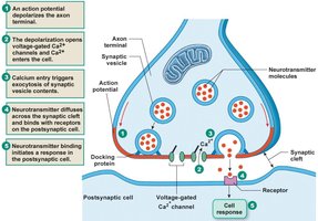
Inactivation of Neurotransmitters
Neurotransmitters are inactivated by enzymatic degradation, reuptake into the presynaptic cell, or diffusion away from the synaptic cleft, ensuring signals are brief and precisely controlled.
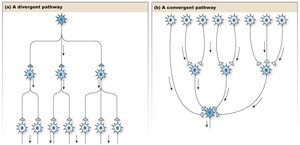
Neuronal Integration: Convergence and Divergence
Neuronal networks integrate information through convergence (many presynaptic neurons synapse onto one postsynaptic neuron) and divergence (one presynaptic neuron branches to affect multiple postsynaptic neurons).
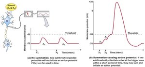
Spatial and Temporal Summation
Summation allows neurons to integrate multiple inputs:
Spatial summation: Simultaneous graded potentials from different locations combine to reach threshold.
Temporal summation: Successive graded potentials from the same location combine if they arrive close enough in time.
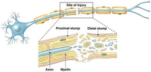
Axon Injury and Regeneration
When an axon is injured, the distal segment degenerates while the proximal segment may survive and regrow, guided by neurotrophic factors. Successful regeneration is more likely in the PNS than in the CNS.
The Central Nervous System
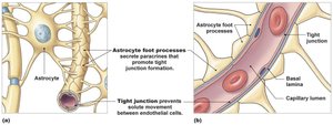
Blood-Brain Barrier
The blood-brain barrier (BBB) is a selective barrier formed by endothelial cells and astrocyte foot processes. It protects the brain from harmful substances while allowing essential nutrients to pass through.
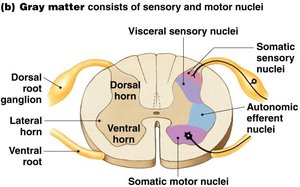
Gray Matter and White Matter
Gray matter consists of neuron cell bodies, dendrites, and axon terminals, while white matter is composed of myelinated axons. In the spinal cord, gray matter is central, and white matter surrounds it.
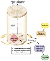
Spinal Cord: Integrating Center
The spinal cord acts as an integrating center for reflexes and transmits information between the brain and the rest of the body. It contains both ascending (sensory) and descending (motor) tracts.
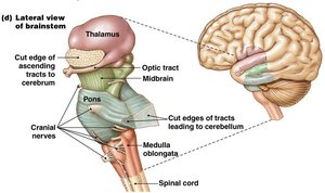
The Brain: Brain Stem and Cerebellum
The brain stem (medulla, pons, midbrain) controls vital involuntary functions such as respiration, heart rate, and reflexes. The cerebellum coordinates movement and processes sensory information.
The Brain: Diencephalon
The diencephalon includes the thalamus (relay and processing center), hypothalamus (homeostasis and endocrine control), pituitary gland (hormone secretion), and pineal gland (melatonin secretion).
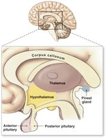
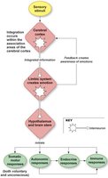
Hypothalamus and Pituitary Gland
The hypothalamus is the major link between the nervous and endocrine systems. It controls the pituitary gland via the infundibulum and regulates many homeostatic functions through hormone secretion.
Gray Matter of the Cerebrum
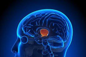
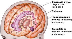
The cerebrum's gray matter includes the cerebral cortex (higher brain functions), basal ganglia (movement control), and limbic system (emotion, learning, memory). The corpus callosum connects the two hemispheres.
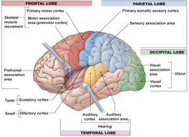
Brain Function: Cerebral Cortex
The cerebral cortex is responsible for sensory perception, voluntary movement, and integration of information. It is divided into sensory, motor, and association areas.
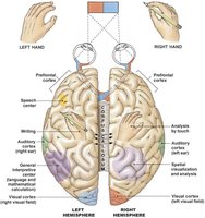
Brain Function: Cerebral Lateralization
The left and right hemispheres of the brain have specialized functions. The left hemisphere is typically associated with language and verbal skills, while the right is associated with spatial skills.
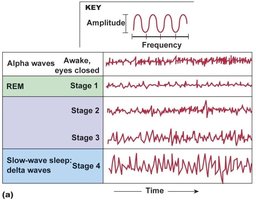
Brain Function: States of Arousal and Sleep
States of arousal, including wakefulness and sleep, are regulated by the reticular activating system. Sleep has two major phases: REM (rapid eye movement) and non-REM (slow-wave) sleep.
Brain Function: Emotion and Moods
The limbic system is the center of emotion and mood regulation. Emotions can influence physiological functions, and mood disorders such as depression are linked to altered synaptic transmission.
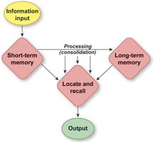
Brain Function: Learning and Memory
Learning can be associative (linking two stimuli) or nonassociative (response to a single stimulus). Memory is classified as short-term or long-term, with consolidation converting short-term to long-term memory.
Reflexive (Implicit) Memory | Declarative (Explicit) Memory | |
|---|---|---|
Recall | Automatic, no conscious attention | Requires conscious attention |
Acquisition | Slow, through repetition | Depends on higher-level thinking |
Includes | Motor skills, procedures | Verbalizable memories |
Demonstration | Procedural | Verbal report |
Brain Function: Language
Language processing involves sensory input, integration in the cerebral cortex, and motor output. Damage to Wernicke's area causes receptive aphasia, while damage to Broca's area causes expressive aphasia.
Brain Function: Personality and Disorders
Personality is shaped by both genetics and experience. Disorders such as schizophrenia have both genetic and environmental components.
