 Back
BackNeurophysiology: Membrane Potentials, Action Potentials, and Synaptic Transmission
Study Guide - Smart Notes
 Tailored notes based on your materials, expanded with key definitions, examples, and context.
Neurophysiology
Resting Membrane Potential (RMP)
The resting membrane potential (RMP) is the electrical charge difference across the membrane of a nonconducting neuron, with the outside being positive and the inside negative. This is due to the distribution of ions and the selective permeability of the membrane.
Extracellular Fluid (ECF): Rich in sodium (Na+), some potassium (K+), and chloride ions (Cl-).
Intracellular Fluid (ICF): Rich in potassium (K+), large negatively charged proteins, and phosphates.
Typical RMP Value: -70 mV; the membrane is said to be polarized.
Ion Channels: Large proteins in the membrane act as ion channels, allowing ions to pass in response to stimuli.
Gated Channels: Open in response to chemical (neurotransmitter), mechanical (pressure), or electrical stimuli.
Generation of an Action Potential
An action potential (AP) is a sequence of events that rapidly changes, reverses, and then restores the membrane potential.
Depolarization: Opening of sodium gates causes Na+ influx, making the inside more positive.
Repolarization: After reaching +30 mV, Na+ gates close and K+ gates open, allowing K+ to exit, restoring negativity.
Key Definitions:
Sub-threshold stimulus: Weak stimulus opens few Na+ channels; not enough for AP.
Threshold stimulus: Opens enough Na+ channels for depolarization; threshold is -55 mV.
All-or-none principle: If threshold is reached, AP is generated fully; stronger stimulus does not increase AP size.
Refractory period: Time when neuron cannot generate another AP.
Propagation of Action Potentials
Action potentials travel along axons by two main mechanisms:
Continuous Conduction: Step-by-step depolarization in unmyelinated axons; slow.
Saltatory Conduction: AP jumps between nodes of Ranvier in myelinated axons; fast.
Signal Transmission at Synapses
A synapse is the junction between two neurons or a neuron and an effector (muscle/gland). Most synapses are chemical, involving neurotransmitter release.
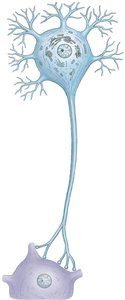
Anatomy of a Chemical Synapse
Presynaptic neuron: Releases neurotransmitter.
Synaptic cleft: Gap between neurons.
Postsynaptic neuron: Receives neurotransmitter.
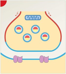
Sequence of Events at the Synapse
The transmission of a signal across a chemical synapse involves several steps:
Action potential arrives at axon terminal, depolarizing the membrane.
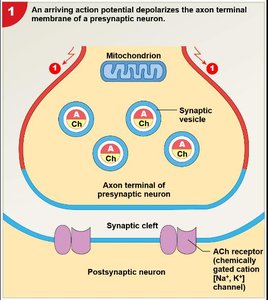
Depolarization opens voltage-gated Ca2+ channels; Ca2+ enters terminal, causing synaptic vesicles to fuse with membrane and release neurotransmitter (e.g., acetylcholine) by exocytosis.
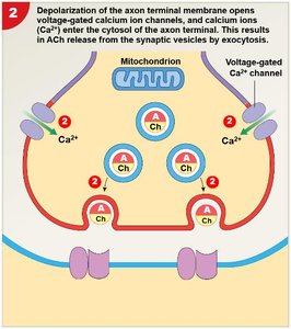
Neurotransmitter diffuses across synaptic cleft, binds to receptors on postsynaptic membrane, opening Na+ and K+ channels and causing graded depolarization.
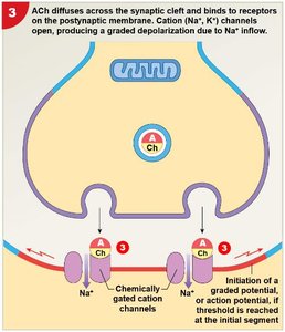
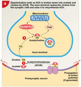
If threshold is reached, a new action potential is generated in the postsynaptic neuron. Acetylcholinesterase breaks down acetylcholine, ending depolarization; choline is reabsorbed and used to resynthesize acetylcholine.
Summary Table: Steps in Synaptic Transmission
Step | Description |
|---|---|
1 | AP arrives at axon terminal, depolarizes membrane |
2 | Ca2+ influx, synaptic vesicle fusion, neurotransmitter release |
3 | Neurotransmitter binds to postsynaptic receptors, channels open |
4 | AP generated in postsynaptic neuron, neurotransmitter breakdown and reuptake |
Key Equations
The Nernst equation can be used to calculate the equilibrium potential for an ion:
Where:
Eion: Equilibrium potential for the ion
R: Gas constant
T: Temperature in Kelvin
z: Charge of the ion
F: Faraday's constant
[ion]outside: Ion concentration outside the cell
[ion]inside: Ion concentration inside the cell
For action potentials, the threshold value is:
Resting membrane potential:
Example: Application in Muscle Contraction
Neurophysiology principles are essential for understanding how nerve impulses trigger muscle contraction. The release of acetylcholine at the neuromuscular junction initiates depolarization in muscle cells, leading to contraction.
