 Back
BackNeurophysiology: Structure and Function of the Nervous System
Study Guide - Smart Notes
 Tailored notes based on your materials, expanded with key definitions, examples, and context.
Neurophysiology: Structure and Function of the Nervous System
Overview of the Nervous System
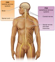
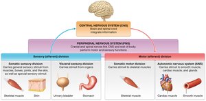
The nervous system is a complex network responsible for coordinating the body's activities by transmitting signals to and from different parts of the body. It is divided into the central nervous system (CNS) and the peripheral nervous system (PNS), each with distinct structures and functions.
Functions of the Nervous System
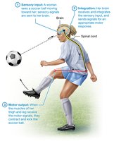
Sensory Input: The nervous system detects changes in the internal and external environment through sensory receptors.
Integration: The CNS processes and interprets sensory input, making decisions about appropriate responses.
Motor Output: The nervous system activates effector organs (muscles and glands) to cause a response.
Divisions of the Nervous System
The nervous system is organized into several divisions based on structure and function:
Central Nervous System (CNS): Consists of the brain and spinal cord; responsible for integrating and processing information.
Peripheral Nervous System (PNS): Includes all neural tissue outside the CNS; subdivided into sensory (afferent) and motor (efferent) divisions.
Motor Division: Further divided into the somatic nervous system (controls skeletal muscles) and the autonomic nervous system (controls smooth muscle, cardiac muscle, and glands).
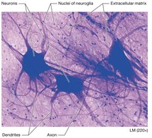
Neurons: Structure and Classification
Anatomy of a Neuron
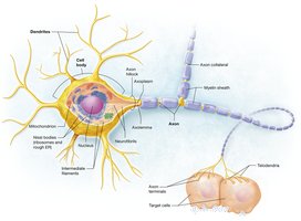
Neurons are the functional units of the nervous system, specialized for the transmission of electrical signals. Each neuron consists of several key anatomical features:
Cell Body (Soma): Contains the nucleus and organelles; responsible for metabolic activities.
Dendrites: Receive incoming signals from other neurons.
Axon: Conducts electrical impulses away from the cell body toward other neurons or effectors.
Axon Hillock: The region where the axon originates; important for action potential initiation.
Myelin Sheath: Insulating layer that increases the speed of signal transmission.
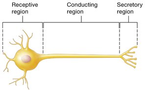
Functional Regions of Neurons
Neurons can be divided into three functional regions based on their roles in signal transmission:
Receptive Region: Includes dendrites and cell body; receives input.
Conducting Region: The axon; transmits action potentials.
Secretory Region: Axon terminals; release neurotransmitters to communicate with other cells.

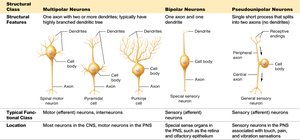
Classification of Neurons
Neurons are classified based on structure and function:
Sensory (Afferent) Neurons: Transmit sensory information toward the CNS.
Motor (Efferent) Neurons: Carry commands from the CNS to muscles and glands.
Interneurons: Connect neurons within the CNS and integrate information.
Structural Type | Features | Function | Location |
|---|---|---|---|
Multipolar | Many dendrites, one axon | Motor, interneurons | CNS, motor neurons in PNS |
Bipolar | One dendrite, one axon | Special sensory (e.g., retina) | Special sense organs |
Pseudounipolar | Single process splits into two | Sensory (afferent) | Sensory neurons in PNS |
Terminology
Nuclei: Clusters of neuron cell bodies in the CNS.
Ganglia: Clusters of neuron cell bodies in the PNS.
Tracts: Bundles of axons in the CNS.
Nerves: Bundles of axons in the PNS.
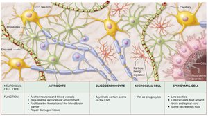
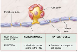
Neuroglia (Glial Cells)
Types and Functions of Glial Cells
Glial cells support, protect, and nourish neurons. They are more numerous than neurons and can divide throughout life.
Astrocytes (CNS): Anchor neurons, regulate the extracellular environment, form the blood-brain barrier, and repair tissue.
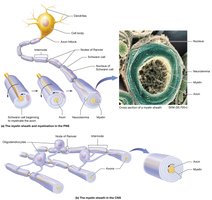
Oligodendrocytes (CNS): Myelinate axons in the CNS.
Microglia (CNS): Act as phagocytes, removing debris and pathogens.
Ependymal Cells (CNS): Line ventricles and produce cerebrospinal fluid.
Schwann Cells (PNS): Myelinate axons in the PNS.
Satellite Cells (PNS): Support cell bodies in ganglia.
Myelin Sheath and Myelination
The myelin sheath is a multilayered covering formed by glial cells (oligodendrocytes in the CNS, Schwann cells in the PNS) that insulates axons and increases the speed of electrical signal transmission. Myelinated regions appear as white matter, while unmyelinated regions (cell bodies, dendrites) are gray matter.

Membrane Potentials and Ion Channels
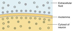
Resting Membrane Potential
The resting membrane potential is the electrical potential difference across the plasma membrane of a neuron at rest, typically about -70 mV. This is maintained by leak channels (especially potassium) and the sodium-potassium pump, resulting in a polarized membrane.
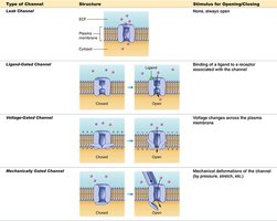
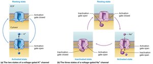
Ion Channels in Neurons
Neurons contain several types of ion channels that contribute to their excitability:
Type of Channel | Structure | Stimulus for Opening/Closing |
|---|---|---|
Leak Channel | Always open | None |
Ligand-Gated Channel | Opens when a chemical binds | Neurotransmitter binding |
Voltage-Gated Channel | Opens with voltage changes | Membrane potential changes |
Mechanically Gated Channel | Opens with mechanical force | Pressure, stretch, etc. |
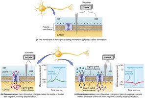
Depolarization and Hyperpolarization
Depolarization occurs when the membrane potential becomes less negative (closer to zero), while hyperpolarization is when it becomes more negative. These changes are critical for the generation and propagation of action potentials.
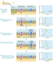
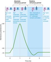
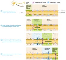
Action Potentials
Generation of Action Potentials
Action potentials are rapid, all-or-nothing electrical impulses that travel along axons. They are triggered when local potentials reach a threshold at the axon hillock, causing voltage-gated sodium and potassium channels to open in sequence.
Events of an Action Potential
Resting state: All voltage-gated channels are closed.
Depolarization: Voltage-gated Na+ channels open, Na+ enters the cell.
Repolarization: Na+ channels inactivate, K+ channels open, K+ exits the cell.
Hyperpolarization: K+ channels remain open briefly, membrane potential becomes more negative than resting.
Return to resting state: Channels reset, resting membrane potential restored.


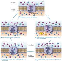
Sodium-Potassium Pump
The sodium-potassium pump (Na+/K+ ATPase) actively transports 3 Na+ ions out of and 2 K+ ions into the neuron, maintaining the resting membrane potential.
Refractory Periods
The refractory period ensures unidirectional propagation of action potentials and limits their frequency:
Absolute Refractory Period: No new action potential can be generated, regardless of stimulus strength.
Relative Refractory Period: A stronger-than-normal stimulus can initiate another action potential.
Local Potentials vs. Action Potentials
Local Potentials: Graded, decremental, reversible, occur in dendrites/cell bodies, can be depolarizing or hyperpolarizing.
Action Potentials: All-or-nothing, nondecremental, irreversible, occur along axons, always depolarizing then repolarizing.
Propagation of Action Potentials
Action potentials propagate along axons by depolarizing adjacent membrane segments. This process is self-propagating and unidirectional due to the refractory period.

Saltatory vs. Continuous Conduction
Saltatory Conduction: Occurs in myelinated axons; action potentials jump between nodes of Ranvier, increasing speed.
Continuous Conduction: Occurs in unmyelinated axons; action potentials propagate along every segment of the membrane.
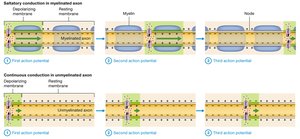
Factors Affecting Conduction Speed
Axon Diameter: Larger diameter = faster conduction due to lower resistance.
Myelination: Myelinated axons conduct signals faster than unmyelinated axons.
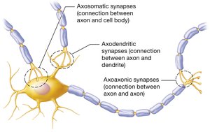
Synapses and Neurotransmission
Synapse Structure and Types
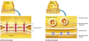
A synapse is the junction where a neuron communicates with its target cell. Types include:
Electrical Synapses: Direct ion flow via gap junctions; rapid and bidirectional.
Chemical Synapses: Use neurotransmitters to transmit signals across a synaptic cleft; unidirectional and modifiable.
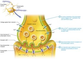
Chemical Synaptic Transmission
An action potential arrives at the axon terminal.
Voltage-gated Ca2+ channels open, Ca2+ enters the terminal.
Neurotransmitter vesicles fuse with the membrane and release contents into the synaptic cleft.
Neurotransmitters bind to receptors on the postsynaptic membrane, generating a postsynaptic potential.
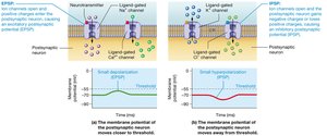
Postsynaptic Potentials
Excitatory Postsynaptic Potential (EPSP): Depolarizes the postsynaptic membrane, increasing the likelihood of an action potential.
Inhibitory Postsynaptic Potential (IPSP): Hyperpolarizes the postsynaptic membrane, decreasing the likelihood of an action potential.
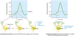
Neural Integration: Temporal and Spatial Summation
Neural integration is the process by which multiple synaptic inputs combine to influence the postsynaptic neuron's activity:
Temporal Summation: Repeated stimulation from a single presynaptic neuron over time.
Spatial Summation: Simultaneous stimulation from multiple presynaptic neurons.
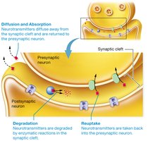
Termination of Synaptic Transmission
Synaptic transmission is terminated by:
Diffusion: Neurotransmitters diffuse away from the synaptic cleft.
Degradation: Enzymes break down neurotransmitters in the cleft.
Reuptake: Neurotransmitters are taken back into the presynaptic neuron for reuse.
Major Neurotransmitters and Their Functions
Acetylcholine: Involved in muscle contraction at the neuromuscular junction.
Norepinephrine: Regulates heart rate, blood pressure, and alertness.
Epinephrine: Similar functions to norepinephrine.
Dopamine: Modulates movement, emotion, and motivation.
Serotonin: Influences mood, attention, and cognition.
Additional info: For a more comprehensive overview, see Table 11.3 in your textbook.
