 Back
Organ Systems, Anatomical Terms, and Body Organization: Step-by-Step Study Guidance
Study Guide - Smart Notes
 Tailored notes based on your materials, expanded with key definitions, examples, and context.
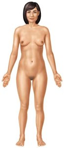
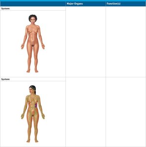
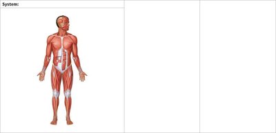
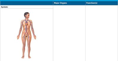
Q1. Identify and name the organ systems shown in the figures. For each system, list its major organs and describe its main functions.
Background
Topic: Human Organ Systems
This question tests your ability to recognize the major organ systems of the human body, identify their key organs, and explain their primary functions. This is foundational knowledge for anatomy and physiology (ANP) courses.
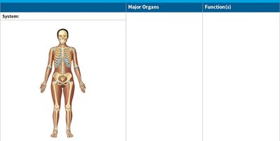
Key Terms and Concepts:
Organ System: A group of organs that work together to perform one or more functions.
Major Organs: The primary anatomical structures that make up each system (e.g., heart in the cardiovascular system).
Function: The main physiological roles of each system (e.g., movement, support, transport of substances).
Step-by-Step Guidance
Examine each figure and note the distinguishing features (e.g., visible muscles, bones, blood vessels, or nerves) to determine which organ system is represented.
For each system, list the major organs you can identify in the diagram (e.g., for the skeletal system: skull, ribs, femur, etc.).
Recall the main function(s) of each system. For example, the skeletal system provides support and protection, while the muscular system enables movement.
Fill in the table for each system with the system name, its major organs, and its primary functions.
Try solving on your own before revealing the answer!
Q2. Describe the anatomical positions and directional terms as shown in the figure.
Background
Topic: Anatomical Position and Directional Terms
This question assesses your understanding of the standard anatomical position and the terminology used to describe locations and directions on the human body.
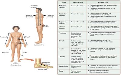
Key Terms and Concepts:
Anatomical Position: The standard reference position for the body in the study of anatomy (standing upright, facing forward, arms at sides, palms facing forward).
Directional Terms: Words used to describe the locations of structures relative to other structures or locations in the body (e.g., anterior, posterior, superior, inferior, medial, lateral, proximal, distal, superficial, deep).
Step-by-Step Guidance
Review the definition of the anatomical position and visualize or describe it in your own words.
Study the directional terms in the figure and match each term to its definition and example (e.g., "anterior" means toward the front).
Practice using these terms to describe the location of one body part relative to another (e.g., the nose is superior to the mouth).
Try solving on your own before revealing the answer!
Q3. Identify the regional terms of the body as shown in the figure.
Background
Topic: Regional Terms of the Human Body
This question tests your ability to recognize and name the specific regions of the body using anatomical terminology.
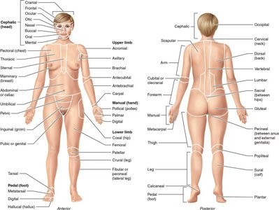
Key Terms and Concepts:
Regional Terms: Specific names given to areas of the body to improve clarity and precision in anatomical description (e.g., brachial for arm, femoral for thigh).
Step-by-Step Guidance
Examine the figure and locate the labeled regions on the anterior and posterior views of the body.
Match each regional term to its corresponding body part (e.g., "axillary" refers to the armpit region).
Practice naming regions for both the upper and lower limbs, as well as the trunk and head.
Try solving on your own before revealing the answer!
Q4. Know the planes of section as shown in the figure.
Background
Topic: Anatomical Planes
This question evaluates your understanding of the three main anatomical planes used to divide the body for study and imaging.
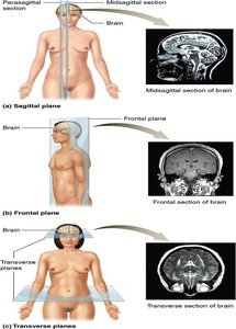
Key Terms and Concepts:
Sagittal Plane: Divides the body into left and right parts (midsagittal is exactly in the middle).
Frontal (Coronal) Plane: Divides the body into anterior (front) and posterior (back) parts.
Transverse (Horizontal) Plane: Divides the body into superior (upper) and inferior (lower) parts.
Step-by-Step Guidance
Identify each plane in the figure and note how it divides the body.
Relate each plane to common medical imaging techniques (e.g., MRI, CT scans).
Practice describing how a section through each plane would appear.
Try solving on your own before revealing the answer!
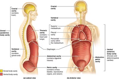
Q5. Know the major body cavities as shown in the figure.
Background
Topic: Body Cavities
This question tests your knowledge of the major cavities in the human body and the organs they contain.
Key Terms and Concepts:
Dorsal Body Cavity: Contains the cranial and vertebral cavities (brain and spinal cord).
Ventral Body Cavity: Contains the thoracic and abdominopelvic cavities (heart, lungs, digestive organs, etc.).
Step-by-Step Guidance
Identify the dorsal and ventral body cavities in the figure.
List the major organs found in each cavity.
Practice describing the boundaries and subdivisions of each cavity (e.g., thoracic cavity contains pleural and pericardial cavities).
Try solving on your own before revealing the answer!
