 Back
BackOrganization and Functional Anatomy of the Cerebrum and Cranial Nerves
Study Guide - Smart Notes
 Tailored notes based on your materials, expanded with key definitions, examples, and context.
Organization of the Cerebrum
Overview of the Cerebrum
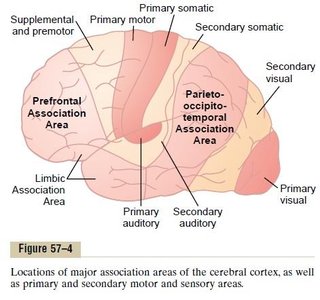
The cerebrum is the largest part of the brain and is responsible for all conscious thought and intellectual functions. Its outer layer, the cerebral cortex, is composed of gray matter (neuronal cell bodies). The characteristic wrinkles (gyri and sulci) increase the surface area, allowing for a greater number of neurons and enhanced processing power. The longitudinal fissure separates the two cerebral hemispheres, each of which is divided into four lobes: frontal, parietal, occipital, and temporal.
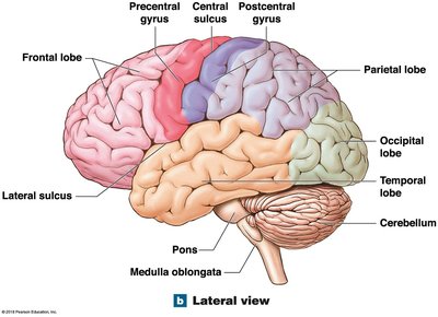
Frontal and Parietal Lobes
The frontal lobe is involved in voluntary motor functions, planning, reasoning, and speech production. Key regions include:
Precentral gyrus: Primary motor cortex, initiates voluntary movements.
Premotor cortex: Plans and coordinates complex movements.
Prefrontal cortex: Involved in executive functions, decision-making, and personality.
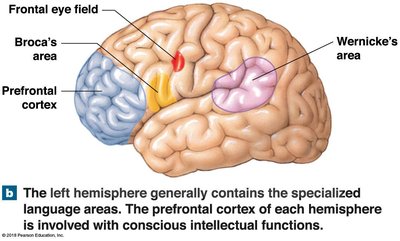
Broca’s area: Critical for speech production.
The central sulcus is a deep groove separating the frontal and parietal lobes. The parietal lobe contains:
Postcentral gyrus: Primary somatosensory cortex, processes touch, pressure, pain, and temperature.
Regions for language processing and somatosensory association.
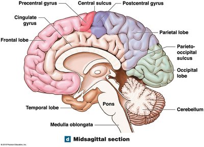
Occipital and Temporal Lobes
The parieto-occipital sulcus separates the parietal and occipital lobes. The occipital lobe is the center for visual processing, containing the primary visual cortex and visual association area. The lateral sulcus separates the parietal/frontal lobes from the temporal lobe. The temporal lobe is responsible for auditory and olfactory processing, containing the primary auditory cortex, auditory association area, and olfactory cortex.
Motor and Sensory Areas of the Cerebral Cortex
Primary Motor and Sensory Cortices
Specific regions of the cortex are dedicated to processing different types of sensory and motor information:
Primary motor cortex (precentral gyrus, frontal lobe): Controls voluntary movements.
Primary somatosensory cortex (postcentral gyrus, parietal lobe): Receives tactile information.
Visual cortex (occipital lobe): Processes visual input.
Auditory cortex (temporal lobe): Processes sound.
Olfactory cortex (temporal lobe): Processes smell.
Gustatory cortex (frontal lobe): Processes taste.
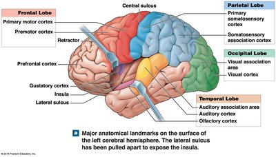
Association Areas
Association areas interpret incoming sensory data and coordinate responses. They include:
Premotor cortex (somatic motor association area): Plans movements.
Visual association area: Interprets visual input.
Auditory association area: Interprets sounds.
Somatosensory association area: Interprets tactile information.

Integrative Areas
Integrative areas are higher-order regions that combine multiple types of sensory input to make complex decisions and direct behavior. These areas are essential for advanced cognitive functions such as problem-solving and planning.
Specialized Language Areas
Wernicke’s and Broca’s Areas
Language processing is localized to specialized regions:
Wernicke’s area: Integrates sensory information and is essential for language comprehension. Damage impairs understanding of language (Wernicke’s aphasia).
Broca’s area: Involved in speech production. Damage impairs the ability to speak (Broca’s aphasia).
Dyslexia: A disorder affecting comprehension and use of written words.
Aphasia: Any disorder affecting the ability to speak or read.
Types of Aphasia
Broca’s aphasia: Difficulty speaking, but comprehension is intact.
Wernicke’s aphasia: Fluent but nonsensical speech; comprehension is impaired.
Global aphasia: Severe impairment in speaking, reading, and understanding speech.
Prefrontal Cortex
Functions of the Prefrontal Cortex
The prefrontal cortex is responsible for abstract intellectual functions, such as predicting consequences, understanding time relationships, making executive decisions, planning for the future, and personality. Damage or surgical alteration (e.g., prefrontal lobotomy) can profoundly affect behavior and personality.

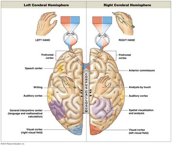
Lateralization of Cerebral Function
Hemispheric Specialization
The two cerebral hemispheres are connected by the corpus callosum and exhibit functional specialization:
Left Hemisphere | Right Hemisphere |
|---|---|
Language, speech, analytical thinking, math/science, logic, detail-oriented | Adjusting to sensory environment, facial recognition, spatial arrangements, emotional context, intuition, creativity |
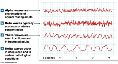
EEGs and Disrupted Brain Physiology
Electroencephalogram (EEG) and Brain Waves
An EEG measures the brain’s electrical activity. Different brain states are associated with characteristic waveforms:
Alpha waves: Awake but relaxed, eyes closed.
Beta waves: Intense concentration, stress, or tension.
Theta waves: Seen in children and frustrated adults.
Delta waves: Deep sleep or certain pathological conditions.
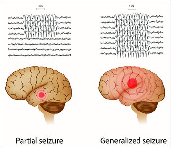
Seizures and Epilepsy
A seizure is caused by excessive synchronous neural activity. Effects depend on the location and spread of abnormal signals. Epilepsy is diagnosed when a person has two or more unprovoked seizures. Febrile seizures are common in children and are triggered by fever.
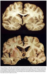
Concussion and Traumatic Brain Injury (TBI)
A concussion is the most common type of TBI, resulting from the brain impacting the inside of the skull. Symptoms may be delayed and can include inflammation, excitotoxicity, and axonal injury. Repeated concussions can lead to Chronic Traumatic Encephalopathy (CTE), a progressive loss of brain function seen in athletes exposed to repeated head trauma.
Cranial Nerves
Olfactory and Optic Nerves
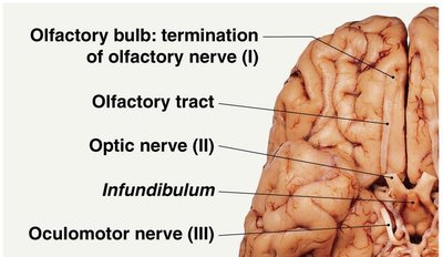
Olfactory nerve (CN I): Sensory only (smell); synapses with the olfactory bulb, whose axons form the olfactory tract.
Optic nerve (CN II): Sensory only (vision); crosses at the optic chiasm and continues as the optic tract.
Control of Eye Movements
Oculomotor nerve (CN III)
Trochlear nerve (CN IV)
Abducens nerve (CN VI)
These nerves control most of the eye’s movements.
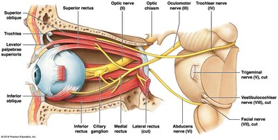
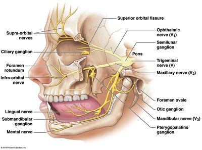
Trigeminal Nerve (CN V)
The trigeminal nerve is a mixed nerve (sensory and motor) with three major divisions:
Ophthalmic (V1)
Maxillary (V2)
Mandibular (V3)
It is involved in facial sensation and mastication, and plays a role in migraine pathophysiology.
Facial and Vestibulocochlear Nerves
Facial nerve (CN VII): Mixed; controls facial muscles and taste sensations. Inflammation can cause Bell’s palsy (facial paralysis).
Vestibulocochlear nerve (CN VIII): Sensory only; carries information for hearing (cochlear branch) and balance (vestibular branch).
Glossopharyngeal and Vagus Nerves
Glossopharyngeal nerve (CN IX): Mixed; innervates the tongue, pharynx, and carotid arteries.
Vagus nerve (CN X): Mixed; widely distributed in the thorax and abdomen, vital for autonomic control of visceral functions.
Accessory and Hypoglossal Nerves
Accessory nerve (CN XI): Motor only; controls muscles of the neck and upper back.
Hypoglossal nerve (CN XII): Motor only; controls tongue movements.
