 Back
BackOrganization and Structure of the Nervous System and Spinal Cord
Study Guide - Smart Notes
 Tailored notes based on your materials, expanded with key definitions, examples, and context.
Nervous System Organization
Overview of Nervous System Function
The nervous system is a highly organized network responsible for coordinating body activities, processing sensory information, and generating motor responses. Most nervous system activity occurs automatically, with conscious awareness involved in only a small portion of daily functions.
Central Nervous System (CNS): Consists of the brain and spinal cord; responsible for processing and integrating information.
Peripheral Nervous System (PNS): Composed of nervous tissue outside the CNS, including nerves and ganglia; transmits sensory and motor signals.
Autonomic vs. Somatic Divisions: The autonomic nervous system (ANS) controls involuntary functions (e.g., heart rate), while the somatic nervous system (SNS) controls voluntary movements (e.g., skeletal muscle contraction).
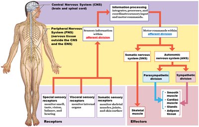
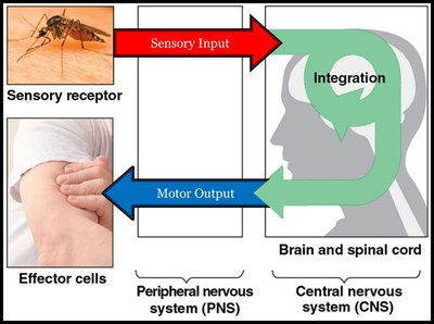
Functional Pathways: Afferent and Efferent Divisions
Sensory Input and Motor Output
Sensory (afferent) pathways carry information from receptors to the CNS, while motor (efferent) pathways transmit commands from the CNS to effectors (muscles and glands).
Afferent Division: Transmits sensory information from receptors (e.g., skin, muscles, organs) to the CNS.
Efferent Division: Carries motor commands from the CNS to effectors (e.g., skeletal muscle, cardiac muscle, glands).
Integration: The CNS processes sensory input and determines appropriate responses.
Spinal Cord Structure and Organization
Gross Anatomy of the Spinal Cord
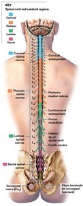
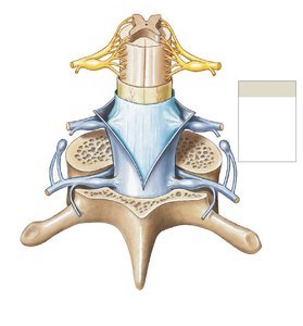
The spinal cord is a cylindrical structure extending from the brainstem to the lower back, protected by vertebrae and meninges. It is divided into cervical, thoracic, lumbar, sacral, and coccygeal regions, each giving rise to spinal nerves that innervate specific body regions.
Cervical and Lumbosacral Enlargements: Regions where nerves serving the limbs arise.
Conus Medullaris: The tapered end of the spinal cord.
Cauda Equina: A bundle of spinal nerve roots extending beyond the conus medullaris.
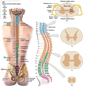
Cross-Sectional Anatomy of the Spinal Cord
In cross-section, the spinal cord displays central gray matter (containing neuron cell bodies) surrounded by white matter (containing myelinated axons). The gray matter is organized into horns, and the white matter into columns.
Gray Matter: Contains sensory and motor nuclei; organized into anterior, posterior, and lateral horns.
White Matter: Contains ascending (sensory) and descending (motor) tracts; organized into anterior, posterior, and lateral columns.
Central Canal: A small channel in the center of the gray matter, containing cerebrospinal fluid (CSF).
Spinal Meninges and Associated Spaces
Protective Layers of the Spinal Cord
The spinal cord is protected by three connective tissue membranes called meninges: dura mater, arachnoid mater, and pia mater. These layers provide physical stability and shock absorption.
Dura Mater: Outermost, tough layer; forms a protective sheath around the spinal cord.
Arachnoid Mater: Middle, weblike layer; contains the subarachnoid space filled with CSF.
Pia Mater: Innermost, delicate layer; adheres closely to the spinal cord surface.
Epidural Space: Space between vertebrae and dura mater; contains adipose and loose connective tissue.
Subarachnoid Space: Space between arachnoid and pia mater; contains CSF, which cushions and nourishes the spinal cord.
Peripheral Nerves and Connective Tissue Wrappings
Structure of Peripheral Nerves
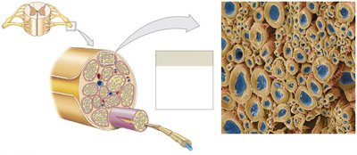
Peripheral nerves are bundles of axons surrounded by connective tissue layers that provide support and protection.
Epineurium: Outermost layer, encloses the entire nerve.
Perineurium: Surrounds bundles of axons called fascicles.
Endoneurium: Surrounds individual axons within a fascicle.
Schwann Cells: Produce myelin sheath around peripheral axons, increasing conduction speed.
Functional Organization of Gray Matter
Nuclei and Horns of the Spinal Cord
Gray matter in the spinal cord is organized into nuclei (clusters of neuron cell bodies) and horns (projections of gray matter). The location of a nucleus determines which body part it controls.
Posterior (Dorsal) Horn: Contains sensory nuclei receiving input from sensory neurons.
Anterior (Ventral) Horn: Contains motor nuclei sending output to skeletal muscles.
Lateral Horn: Contains autonomic motor nuclei (present in thoracic and upper lumbar regions).
Commissures: Regions where axons cross from one side of the spinal cord to the other (anterior and posterior gray commissures).
Spinal Reflexes
Reflex Arc and Types of Reflexes
Reflexes are rapid, automatic responses to specific stimuli. The basic pathway is called a reflex arc, which includes a receptor, sensory neuron, integration center, motor neuron, and effector.
Spinal Reflexes: Processed in the spinal cord (e.g., withdrawal reflex).
Cranial Reflexes: Processed in the brain (e.g., blinking).
Somatic Reflexes: Involve skeletal muscles.
Visceral (Autonomic) Reflexes: Involve smooth muscle, cardiac muscle, or glands.
Examples of Spinal Reflexes
Stretch Reflex: Maintains muscle length and tone (e.g., knee-jerk reflex).
Withdrawal Reflex: Moves a body part away from a painful stimulus.
Crossed Extensor Reflex: Involves contralateral activation of muscles to maintain balance during withdrawal.
Summary Table: Organization of the Spinal Cord
Region | Gray Matter | White Matter | Associated Function | |
|---|---|---|---|---|
Posterior Horn | Sensory nuclei | Posterior columns | Sensory processing | |
Anterior Horn | Motor nuclei | Anterior columns | Motor output to skeletal muscle | |
Lateral Horn | Autonomic motor nuclei | Lateral columns | Autonomic (visceral) motor control |
Additional info: The spinal cord is essential for both transmitting information between the brain and body and for mediating reflexes that do not require conscious thought. The organization of gray and white matter allows for efficient processing and relay of sensory and motor signals.
