 Back
BackOverview of Major Human Muscles: Structure and Function
Study Guide - Smart Notes
 Tailored notes based on your materials, expanded with key definitions, examples, and context.
Muscles of the Human Body
The muscular system is essential for movement, posture, and various bodily functions. This guide provides an overview of the major muscle groups, their anatomical locations, and their primary functions, with visual references for each region.
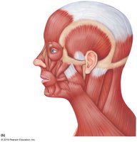
Head and Neck Muscles
The muscles of the head and neck are responsible for facial expressions, mastication (chewing), head movement, and supporting the skull. Key muscles include the occipitofrontalis, temporalis, masseter, sternocleidomastoid, and trapezius.
Occipitofrontalis: Raises the eyebrows and wrinkles the forehead.
Temporalis and Masseter: Elevate the mandible for chewing.
Sternocleidomastoid: Rotates and flexes the neck.
Trapezius: Moves the scapula and supports the arm.
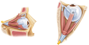
Eye Muscles
The extrinsic eye muscles control the movement of the eyeball and are crucial for vision. There are six main muscles: superior rectus, inferior rectus, lateral rectus, medial rectus, superior oblique, and inferior oblique.
Rectus muscles: Move the eye up, down, and side-to-side.
Oblique muscles: Rotate the eye and assist in complex movements.
Innervation: Most are innervated by the oculomotor nerve (CN III), except the lateral rectus (abducens, CN VI) and superior oblique (trochlear, CN IV).
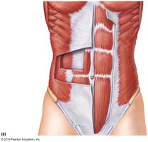
Abdominal Muscles
The abdominal muscles support the trunk, allow movement, and hold organs in place. The main muscles are the rectus abdominis, external oblique, internal oblique, and transversus abdominis.
Rectus abdominis: Flexes the vertebral column and compresses the abdomen.
Obliques: Rotate and laterally flex the trunk.
Transversus abdominis: Compresses abdominal contents.
Linea alba: A fibrous structure running down the midline of the abdomen.
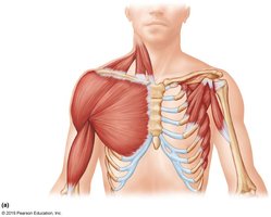
Upper Torso Muscles
The upper torso muscles are involved in movements of the shoulder, arm, and chest. Major muscles include the pectoralis major, deltoid, serratus anterior, and intercostal muscles.
Pectoralis major: Adducts and medially rotates the arm.
Deltoid: Abducts the arm.
Serratus anterior: Protracts the scapula.
Intercostal muscles: Assist in respiration by moving the rib cage.
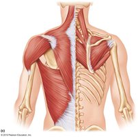
Back Muscles
The back muscles stabilize and move the vertebral column and shoulder blades. Key muscles include the trapezius, latissimus dorsi, rhomboids, and erector spinae group.
Trapezius: Elevates, retracts, and rotates the scapula.
Latissimus dorsi: Extends, adducts, and medially rotates the arm.
Rhomboids: Retract the scapula.
Erector spinae: Extends and laterally flexes the vertebral column.
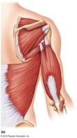
Back and Arm Muscles
This region includes muscles that connect the back to the upper arm, facilitating arm movement and stabilization. Important muscles are the deltoid, infraspinatus, teres major, and triceps brachii.
Deltoid: Main abductor of the arm.
Infraspinatus and teres major: Rotate the arm laterally and medially, respectively.
Triceps brachii: Extends the forearm at the elbow.
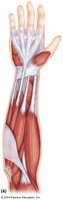
Forearm Muscles
The forearm muscles control movements of the wrist, hand, and fingers. They are divided into flexors (anterior) and extensors (posterior).
Flexor group: Flex the wrist and fingers.
Extensor group: Extend the wrist and fingers.
Pronator and supinator muscles: Rotate the forearm.
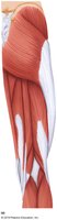
Anterior Thigh Muscles
The anterior thigh muscles are primarily responsible for extending the knee and flexing the hip. The main group is the quadriceps femoris, which includes four muscles: rectus femoris, vastus lateralis, vastus medialis, and vastus intermedius.
Quadriceps femoris: Extends the knee.
Sartorius: Flexes, abducts, and laterally rotates the thigh.
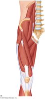
Posterior Thigh Muscles
The posterior thigh muscles, known as the hamstrings, are responsible for flexing the knee and extending the hip. The group includes the biceps femoris, semitendinosus, and semimembranosus.
Hamstrings: Flex the knee and extend the hip.
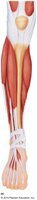
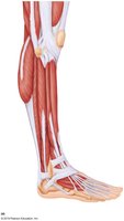
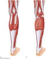
Lower Leg Muscles
The lower leg muscles are divided into anterior, lateral, and posterior compartments, each with specific functions in foot and toe movement.
Tibialis anterior: Dorsiflexes the foot.
Gastrocnemius and soleus: Plantarflex the foot.
Fibularis (peroneus) muscles: Evert the foot.
Flexor and extensor digitorum muscles: Move the toes.
Additional info:
Muscle actions are often paired (agonist/antagonist) to allow for smooth and controlled movements.
Understanding muscle anatomy is essential for clinical assessment, injury prevention, and rehabilitation.
