 Back
BackOverview of Skeletal System and Joints in Human Anatomy
Study Guide - Smart Notes
 Tailored notes based on your materials, expanded with key definitions, examples, and context.
Skeletal System Overview
Introduction to the Skeletal System
The skeletal system provides the structural framework for the human body, protects vital organs, facilitates movement, stores minerals, and houses bone marrow for blood cell production. It is composed of bones, cartilage, ligaments, and joints.
Bones: Rigid organs that form the skeleton and support the body.
Cartilage: Flexible connective tissue found in joints, ear, nose, and other structures.
Ligaments: Bands of dense connective tissue connecting bones to other bones.
Joints: Articulations where two or more bones meet, allowing for movement and flexibility.
Axial and Appendicular Skeleton
Axial Skeleton
The axial skeleton forms the central axis of the body and includes the skull, vertebral column, and thoracic cage.
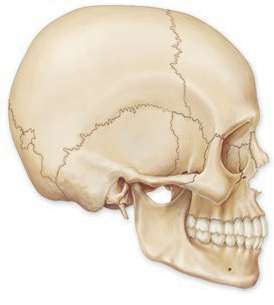
Skull: Protects the brain and forms the structure of the face.
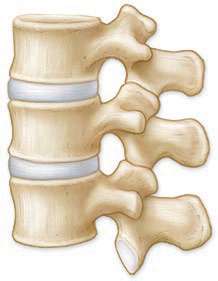
Vertebral Column: Supports the body and protects the spinal cord.
Thoracic Cage: Composed of ribs and sternum, protects the heart and lungs.
Appendicular Skeleton
The appendicular skeleton consists of the bones of the limbs and girdles that attach them to the axial skeleton.
Pectoral Girdle: Clavicle and scapula, attaching the upper limbs to the trunk.
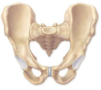
Pelvic Girdle: Hip bones, attaching the lower limbs to the trunk.
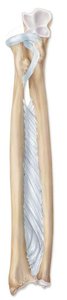
Upper and Lower Limbs: Bones of the arms, hands, legs, and feet.

Joints (Articulations)
Classification of Joints
Joints are classified based on their structure and function. They allow for varying degrees of movement and stability.
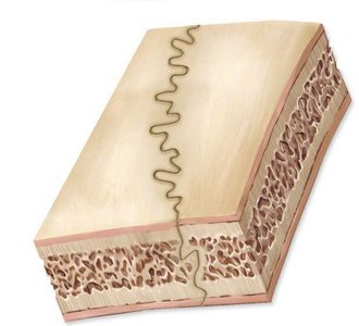
Fibrous Joints: Bones joined by dense connective tissue; mostly immovable (e.g., sutures of the skull).
Cartilaginous Joints: Bones joined by cartilage; allow limited movement (e.g., intervertebral discs, pubic symphysis).
Synovial Joints: Freely movable joints with a synovial cavity (e.g., shoulder, knee, elbow).


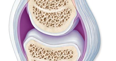
Structure of Synovial Joints
Synovial joints are the most common and movable type of joint in the body. They are characterized by the presence of a synovial cavity filled with fluid, articular cartilage, and a joint capsule.
Articular Cartilage: Covers the ends of bones, reducing friction and absorbing shock.
Synovial Membrane: Lines the joint capsule and secretes synovial fluid for lubrication.
Joint Capsule: Encloses the joint cavity and provides stability.
Ligaments: Strengthen and support the joint.

Specialized Structures in Bones and Joints
Growth Plate (Epiphyseal Plate)
The epiphyseal plate is a region of cartilage found in growing bones, responsible for longitudinal bone growth during childhood and adolescence.
Location: Between the epiphysis and diaphysis of long bones.
Function: Allows bones to lengthen until adulthood, when it ossifies and becomes the epiphyseal line.
Bone Structure and Marrow
Bones are composed of compact and spongy bone tissue, with marrow cavities containing red or yellow marrow.
Compact Bone: Dense outer layer providing strength.
Spongy Bone: Porous inner layer containing trabeculae and marrow spaces.
Red Marrow: Site of blood cell production (hematopoiesis).
Yellow Marrow: Stores fat.
Examples of Joints in the Human Body
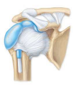
Shoulder Joint (Glenohumeral Joint)
The shoulder joint is a ball-and-socket synovial joint, allowing for a wide range of motion in the upper limb.
Articulating Bones: Humerus and scapula.
Movements: Flexion, extension, abduction, adduction, rotation, and circumduction.
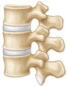
Vertebral Joints
Vertebrae are joined by intervertebral discs (cartilaginous joints) and facet joints (synovial joints), allowing for flexibility and movement of the spine.
Intervertebral Discs: Absorb shock and allow slight movement between vertebrae.
Facet Joints: Allow for gliding movements between vertebrae.

Pelvic Joints
The pelvic girdle includes the sacroiliac joints (between sacrum and ilium) and the pubic symphysis (between pubic bones), providing stability and support for the lower body.
Sacroiliac Joint: Slightly movable joint between sacrum and ilium.
Pubic Symphysis: Cartilaginous joint uniting the left and right pubic bones.
Summary Table: Types of Joints
Type of Joint | Structural Features | Example | Movement |
|---|---|---|---|
Fibrous | Dense connective tissue, no cavity | Sutures of skull | Immovable |
Cartilaginous | Cartilage, no cavity | Intervertebral discs, pubic symphysis | Slightly movable |
Synovial | Synovial cavity, articular cartilage, capsule | Shoulder, knee, elbow | Freely movable |
Conclusion
The skeletal system and its joints are essential for support, movement, and protection of the human body. Understanding the structure and function of bones and joints is fundamental for further study in anatomy and physiology.
