 Back
BackPeripheral and Autonomic Nervous System: Structure, Function, and Special Senses
Study Guide - Smart Notes
 Tailored notes based on your materials, expanded with key definitions, examples, and context.
Peripheral Nervous System (PNS)
PNS Structures
The Peripheral Nervous System (PNS) connects the central nervous system (CNS) to limbs and organs, facilitating communication between the brain, spinal cord, and the rest of the body. It consists of spinal nerves, cranial nerves, ganglia, and sensory receptors.
12 Cranial Nerves: Originate from the brain and brainstem; transmit sensory and motor information.
31 Spinal Nerves: Cervical (8 pairs), Thoracic (12 pairs), Lumbar (5 pairs), Sacral (5 pairs), Coccygeal (1 pair).
Plexuses: Networks of axons from anterior rami (except thoracic T2-T12).
Ganglia: Dorsal root (sensory) and autonomic (motor; sympathetic and parasympathetic).
Cranial Nerves: Classification and Function
Sensory Nerves: Olfactory (I), Optic (II), Vestibulocochlear (VIII)
Motor Nerves: Oculomotor (III), Trochlear (IV), Abducens (VI), Accessory (XI), Hypoglossal (XII)
Mixed Nerves: Trigeminal (V), Facial (VII), Glossopharyngeal (IX), Vagus (X)
Examples:
Olfactory Nerve (I): Sense of smell.
Optic Nerve (II): Vision.
Trigeminal Nerve (V): Sensation in face, motor for chewing.
Vagus Nerve (X): Parasympathetic control of heart, lungs, digestive tract.
Plexuses and Major Nerves
Cervical Plexus: Innervates head, neck, shoulders; phrenic nerve controls diaphragm.
Brachial Plexus: Innervates shoulder, upper limbs; includes musculocutaneous, axillary, median, radial, ulnar nerves.
Lumbar Plexus: Innervates abdomen, pelvis, lower limbs.
Sacral Plexus: Innervates pelvis, legs; sciatic nerve is largest.
Coccygeal Plexus: Smallest; anococcygeal nerve innervates coccyx region.
Autonomic Nervous System (ANS) and Homeostasis
Overview and Function
The Autonomic Nervous System (ANS) regulates involuntary functions by innervating smooth muscle, cardiac muscle, and glands. It operates through two main divisions: sympathetic and parasympathetic, maintaining homeostasis and responding to stress.
Efferent Pathway: Consists of two neurons (preganglionic and postganglionic).
Main Integration Center: Hypothalamus.
Sympathetic Division (“Fight or Flight”)
Activated during stress or physical activity.
Effects: Increased heart rate, blood pressure, bronchiole dilation, decreased GI activity, pupil dilation, sweat gland stimulation.
Preganglionic Neurons: Thoracolumbar region.
Ganglia: Sympathetic trunk (close to spinal cord), collateral ganglia.
Rami Communicantes: White (preganglionic to ganglia), Gray (postganglionic to spinal nerves).
Adrenal Medulla: Secretes epinephrine and norepinephrine; acts as hormonal backup.
Parasympathetic Division (“Rest and Digest”)
Activated during rest; promotes energy conservation.
Effects: Decreased heart rate, increased GI activity, pupil constriction, urination.
Preganglionic Neurons: Craniosacral region.
Vagus Nerve (CN X): Provides ~90% of parasympathetic innervation.
Terminal Ganglia: Located near or within target organs.
Control Centers of the ANS
Hypothalamus: Directs autonomic functions.
Brainstem: Relay center for autonomic communication.
Reticular Formation: Regulates skeletal and visceral muscle activity.
Limbic System: Processes emotional stimuli.
Visceral Reflex Arcs
Visceral reflex arcs regulate involuntary responses in smooth muscle, cardiac muscle, and glands. They differ from somatic reflex arcs by having two motor neurons and visceral sensory afferents.
Receptor: Sensory receptor in viscera
Visceral Sensory Neuron: Transmits impulse to CNS
Integration Center: Interneuron or single synapse
Motor Neuron: Preganglionic and postganglionic neurons
Visceral Effector: Smooth muscle, cardiac muscle, or gland
Neurotransmitters and Receptors
Sympathetic Division: Preganglionic fibers release acetylcholine (ACh); postganglionic fibers release norepinephrine, epinephrine, or ACh.
Parasympathetic Division: Both pre- and postganglionic fibers release ACh.
Cholinergic Fibers: Release/respond to ACh (muscarinic and nicotinic receptors).
Adrenergic Fibers: Release norepinephrine and epinephrine (alpha and beta receptors).
Neurotransmitter Receptors
Adrenergic Receptors: Bind norepinephrine/epinephrine; found in sympathetic target organs.
Cholinergic Receptors: Bind acetylcholine; nicotinic (at all ANS ganglia and adrenal medulla), muscarinic (parasympathetic target organs, sweat glands).
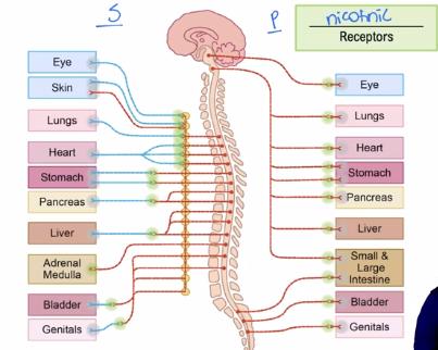
Illustration: Sympathetic vs. Parasympathetic Innervation
The following image visually compares the target organs innervated by the sympathetic (S) and parasympathetic (P) divisions, highlighting the pathways and the presence of nicotinic receptors at ganglia.
The Senses: General and Special
General Senses
General senses are distributed throughout the body and include touch, pain, and proprioception. They rely on various receptor types:
Mechanoreceptors: Respond to physical deformation (touch, pressure, stretch, vibration).
Nociceptors: Respond to painful stimuli (extreme heat, pressure, chemicals).
Proprioceptors: Detect stretch in muscles and tendons, providing awareness of body position.
Exteroceptors: Respond to external stimuli (skin receptors for pressure, pain, temperature).
Sensory Adaptation: Receptors become less responsive to constant, unchanging stimulus.
Transduction: Conversion of a stimulus into a nerve impulse.
Dermatome: Area of skin innervated by a single spinal nerve; useful for locating spinal cord injuries.
Special Senses
Special senses are located in specialized organs in the head and include vision, smell, taste, hearing, and equilibrium.
Vision: Eyes; stimulus is light; perception of brightness and color.
Smell (Olfaction): Olfactory epithelium; stimulus is chemical; perception of odors.
Taste (Gustation): Taste buds; stimulus is chemical; perception of sweet, sour, salty, bitter, umami.
Hearing: Ear and cochlea; stimulus is pressure waves; perception of sound.
Equilibrium: Semicircular canals and vestibule; stimulus is movement and gravity; perception of balance.
Vision: Structure and Function of the Eye
Layers of the Eyeball
Fibrous Layer: Cornea (transparent, allows light entry, pain receptors, avascular), Sclera (white, protects, shapes, anchors muscles, vascular).
Vascular Layer: Iris (regulates light entry), Pupil (hole for light), Ciliary Body (controls lens shape, produces fluid), Choroid (absorbs excess light, blood supply).
Inner Layer: Retina (photoreceptors, nerve cells), Fovea centralis (center of vision, only cones).
Optic Components
Aqueous Humor: Nourishes lens and cornea; buildup can cause glaucoma.
Vitreous Humor: Maintains eye shape; made during fetal development.
Lens: Focuses light; changes in crystallin proteins cause cataracts.
Retina and Photoreceptors
Rods: Grayscale vision, high sensitivity, low acuity, peripheral location.
Cones: Color vision, low sensitivity, high acuity, central location (fovea).
Macula Lutea: Center of vision, mostly cones.
Optic Disc: Blind spot, no photoreceptors.
Light Control and Accommodation
Pupil Constriction: Parasympathetic innervation; bright light, close vision.
Pupil Dilation: Sympathetic innervation; dim light, distant vision.
Accommodation Reflex: Ciliary muscles contract, lens bulges, pupil constricts, eyes converge for near vision.
Presbyopia: Age-related loss of lens flexibility.
Hearing and Balance
Hearing (Auditory)
Receptors: Located in cochlea.
Sequence: Sound waves move basilar membrane → stereocilia bend → K+ channels open → depolarization → neurotransmitter release → action potential in cochlear nerve.
Ear Anatomy: Pinna, external auditory canal, tympanic membrane, ossicles (malleus, incus, stapes), cochlea, semicircular canals, vestibulocochlear nerve.
Balance (Equilibrium)
Receptors: Vestibular apparatus (semicircular canals, vestibule).
Dynamic Equilibrium: Crista ampullaris detects rotational movement.
Static Equilibrium: Maculae detect linear acceleration and head position.
Physiology: Stereocilia bend toward kinocilium → depolarization → increased glutamate release → more action potentials.
Clinical: Balance problems often involve vestibulocochlear nerve (CN VIII).
Taste (Gustation) and Smell (Olfaction)
Taste (Gustation)
Transduction Sequence: Chemical binds to gustatory cell → ion movement → depolarization → Ca²+ channels open → neurotransmitter release → action potential.
Taste Sensations: Sweet, sour, salty, bitter, umami (triggered by amino acids).
Smell (Olfaction)
Organ: Olfactory epithelium.
Clinical: Inability to distinguish odors may indicate olfactory nerve (CN I) damage.
Key Anatomy Identification
Neuron: Dendrites, cell body (soma), axon, axon terminals.
Eye: Cornea, iris, lens, vitreous humor, retina, optic nerve.
Ear: Pinna, external auditory canal, tympanic membrane, ossicles, cochlea, semicircular canals, vestibulocochlear nerve.
Hyperpolarization and Inhibition
Hyperpolarization increases the difficulty of generating an action potential by moving the neuron further from threshold, requiring stronger excitatory input. This is a key mechanism for inhibitory neurotransmitters in the ANS.
Division | Preganglionic Neurotransmitter | Postganglionic Neurotransmitter | Main Receptors |
|---|---|---|---|
Sympathetic | Acetylcholine (ACh) | Norepinephrine, Epinephrine, ACh | Adrenergic (alpha, beta), Cholinergic (nicotinic, muscarinic) |
Parasympathetic | Acetylcholine (ACh) | Acetylcholine (ACh) | Cholinergic (nicotinic, muscarinic) |
Additional info: The included image directly illustrates the pathways and target organs of the sympathetic and parasympathetic divisions, emphasizing the role of nicotinic receptors at ganglia, which is a core concept in ANS physiology.
