 Back
BackPeripheral Nervous System & Reflex Activity Study Guide
Study Guide - Smart Notes
 Tailored notes based on your materials, expanded with key definitions, examples, and context.
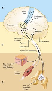
Q1. Identify the three basic levels of neural integration in sensory systems labeled A–C. Refer to Figure 13.2 for help.
Background
Topic: Sensory Processing in the Nervous System
This question tests your understanding of how sensory information is processed and integrated at different levels within the nervous system, from the initial detection of stimuli to perception in the brain.
Key Terms:
Sensory receptors: Specialized cells or structures that detect changes in the environment.
Neural integration: The process by which the nervous system processes and interprets sensory input.
Ascending pathways: Neural pathways that carry sensory information toward the brain.
Cerebral cortex: The region of the brain responsible for conscious perception and interpretation of sensory stimuli.
Step-by-Step Guidance
Examine the diagram (Figure 13.2) and note the three labeled regions: A, B, and C. Each region represents a distinct level of sensory processing.
Recall that sensory integration typically occurs at three levels: the receptor level (where sensory receptors detect stimuli), the circuit level (where information is relayed through ascending pathways), and the perceptual level (where the cerebral cortex interprets the information).
Match each label (A, B, C) to its corresponding level based on the structures shown in the diagram. For example, A may represent the perceptual level (cerebral cortex), B the circuit level (spinal cord, brainstem), and C the receptor level (sensory receptors in skin, muscle, joint).
Use the diagram to identify which structures are involved at each level and how information flows from the periphery to the brain.
Try solving on your own before revealing the answer!
Final Answer:
A: Perceptual level (cerebral cortex) B: Circuit level (spinal cord, brainstem) C: Receptor level (sensory receptors)
These levels represent the flow of sensory information from detection (receptors), through neural circuits (spinal cord/brainstem), to conscious perception (cerebral cortex).
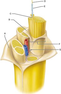
Q2. Name the parts of a nerve labeled A–G. Refer to Figure 13.4 for help.
Background
Topic: Structure of a Nerve
This question assesses your knowledge of the anatomy of a nerve, including its connective tissue coverings and internal organization.
Key Terms:
Axon: The long, slender projection of a neuron that conducts electrical impulses.
Fascicle: A bundle of axons within a nerve.
Endoneurium: Connective tissue surrounding individual axons.
Perineurium: Connective tissue surrounding each fascicle.
Epineurium: Connective tissue surrounding the entire nerve.
Step-by-Step Guidance
Study the diagram (Figure 13.4) and locate the labels A–G. Each label points to a specific structure within the nerve.
Recall the three main connective tissue layers: endoneurium (around individual axons), perineurium (around fascicles), and epineurium (around the whole nerve).
Identify which label corresponds to each layer and to other structures such as blood vessels, fascicles, and axons.
Use your textbook or class notes to match the anatomical terms to the labeled parts in the diagram.
Try solving on your own before revealing the answer!
Final Answer:
A: Epineurium B: Fascicle C: Perineurium D: Axon E: Endoneurium F: Blood vessel G: Myelin sheath
These structures collectively form the organization of a peripheral nerve, allowing for efficient transmission of sensory and motor signals.
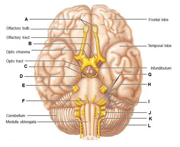
Q3. Provide the name and Roman numeral for each of the cranial nerves indicated (A–L). Refer to Figure 13.6 for help.
Background
Topic: Cranial Nerves
This question tests your ability to identify the cranial nerves by their anatomical location and assign the correct Roman numeral to each.
Key Terms:
Cranial nerves: Twelve pairs of nerves that emerge directly from the brain and innervate structures in the head and neck.
Roman numerals: Used to number the cranial nerves (I–XII).
Step-by-Step Guidance
Examine the diagram (Figure 13.6) and locate the labels A–L. Each label corresponds to a cranial nerve.
Recall the names and Roman numerals of the twelve cranial nerves in order: Olfactory (I), Optic (II), Oculomotor (III), Trochlear (IV), Trigeminal (V), Abducens (VI), Facial (VII), Vestibulocochlear (VIII), Glossopharyngeal (IX), Vagus (X), Accessory (XI), Hypoglossal (XII).
Match each label to the correct nerve based on its anatomical location and pathway shown in the diagram.
Assign the appropriate Roman numeral to each nerve.
Try solving on your own before revealing the answer!
Final Answer:
A: Olfactory (I) B: Optic (II) C: Oculomotor (III) D: Trochlear (IV) E: Trigeminal (V) F: Abducens (VI) G: Facial (VII) H: Vestibulocochlear (VIII) I: Glossopharyngeal (IX) J: Vagus (X) K: Accessory (XI) L: Hypoglossal (XII)
Each cranial nerve is identified by its function and anatomical location, and numbered with a Roman numeral.
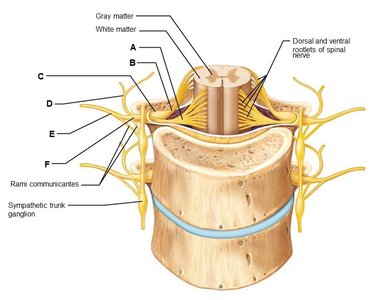
Q4. Name the structures associated with the spinal cord and spinal nerves labeled A–F. Refer to Figure 13.8 for help.
Background
Topic: Spinal Cord and Spinal Nerve Anatomy
This question tests your ability to identify key anatomical structures related to the spinal cord and its associated nerves.
Key Terms:
Gray matter: Region of the spinal cord containing neuron cell bodies.
White matter: Region containing myelinated axons.
Dorsal and ventral roots: Pathways for sensory and motor information.
Rami communicantes: Branches connecting spinal nerves to the sympathetic trunk.
Step-by-Step Guidance
Study the diagram (Figure 13.8) and locate the labels A–F. Each label points to a specific structure associated with the spinal cord and nerves.
Recall the main anatomical features: gray matter, white matter, dorsal and ventral roots, rami communicantes, and sympathetic trunk ganglion.
Match each label to the correct anatomical term based on its position and appearance in the diagram.
Use your textbook or class notes to confirm the identity of each labeled structure.
Try solving on your own before revealing the answer!
Final Answer:
A: Gray matter B: White matter C: Dorsal and ventral rootlets of spinal nerve D: Rami communicantes E: Sympathetic trunk ganglion F: Spinal nerve
These structures are essential for the transmission of sensory and motor signals between the body and the central nervous system.
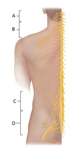
Q5. Name the nerve plexuses (A–D). Refer to Figure 13.7 for help.
Background
Topic: Nerve Plexuses
This question tests your knowledge of the major nerve plexuses in the body and their anatomical locations.
Key Terms:
Nerve plexus: A network of intersecting nerves.
Cervical, brachial, lumbar, and sacral plexuses: The four major plexuses.
Step-by-Step Guidance
Examine the diagram (Figure 13.7) and locate the labels A–D. Each label corresponds to a major nerve plexus.
Recall the four major plexuses: cervical (neck), brachial (shoulder/arm), lumbar (lower back), and sacral (pelvis/leg).
Match each label to the correct plexus based on its anatomical location in the diagram.
Use your textbook or class notes to confirm the identity of each plexus.
Try solving on your own before revealing the answer!
Final Answer:
A: Cervical plexus B: Brachial plexus C: Lumbar plexus D: Sacral plexus
Each plexus serves specific regions of the body, providing motor and sensory innervation.
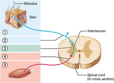
Q6. Complete the table by naming and briefly describing the function of each of the components of a reflex arc indicated 1–5 in the figure below. Refer to Figure 13.15 for help.
Background
Topic: Reflex Arc Components
This question tests your understanding of the basic components of a reflex arc and their roles in producing rapid, automatic responses.
Key Terms:
Reflex arc: The neural pathway involved in a reflex action.
Receptor, sensory neuron, integration center, motor neuron, effector: The five main components.
Step-by-Step Guidance
Study the diagram (Figure 13.15) and locate the numbered components 1–5. Each number corresponds to a specific part of the reflex arc.
Recall the sequence of events in a reflex arc: stimulus detected by receptor, signal transmitted by sensory neuron, processed in integration center, response sent by motor neuron, and action carried out by effector.
Match each number to the correct component and briefly describe its function.
Use your textbook or class notes to confirm the identity and function of each component.
Try solving on your own before revealing the answer!
Final Answer:
1: Receptor – detects the stimulus 2: Sensory neuron – transmits the signal to the CNS 3: Integration center – processes the information 4: Motor neuron – carries the response signal 5: Effector – produces the response
These components work together to produce rapid, predictable responses to stimuli.
