 Back
Peripheral Nervous System & Reflex Activity: Structured Study Notes
Study Guide - Smart Notes
 Tailored notes based on your materials, expanded with key definitions, examples, and context.
Peripheral Nervous System (PNS) Overview
Structure and Function of the PNS
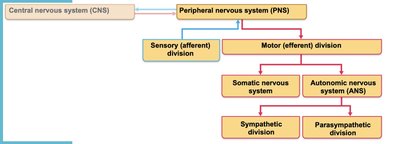
The Peripheral Nervous System (PNS) comprises all neural structures outside the brain and spinal cord. It includes sensory receptors, peripheral nerves, associated ganglia, and motor endings, providing essential links between the central nervous system (CNS) and the external environment.
Sensory receptors: Specialized to respond to environmental changes (stimuli).
Peripheral nerves: Bundles of axons transmitting signals.
Ganglia: Collections of neuron cell bodies associated with nerves.
Motor endings: Structures that activate effectors (muscles/glands).
Sensory Receptors
Classification by Stimulus Type
Sensory receptors are classified based on the type of stimulus they detect:
Mechanoreceptors: Respond to touch, pressure, vibration, stretch, and itch.
Thermoreceptors: Sensitive to temperature changes.
Photoreceptors: Respond to light energy (e.g., retina).
Chemoreceptors: Respond to chemicals (e.g., smell, taste, blood chemistry).
Nociceptors: Sensitive to pain-causing stimuli (e.g., extreme heat/cold, pressure, chemicals).
Classification by Location
Exteroceptors: Respond to stimuli outside the body; located near the body surface; sensitive to touch, pressure, pain, temperature; include special sense organs.
Interoceptors: Respond to stimuli within the body; found in internal viscera and blood vessels; sensitive to chemical changes, stretch, temperature.
Proprioceptors: Respond to degree of stretch; found in skeletal muscles, tendons, joints, ligaments; inform the brain of movements.
Classification by Structural Complexity
Receptors are structurally classified as simple (encapsulated or unencapsulated) or complex (special sense organs).
Simple Receptors: Unencapsulated
Free nerve endings of sensory neurons
Modified free nerve endings (Merkel cells/discs, hair follicle receptors)
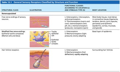
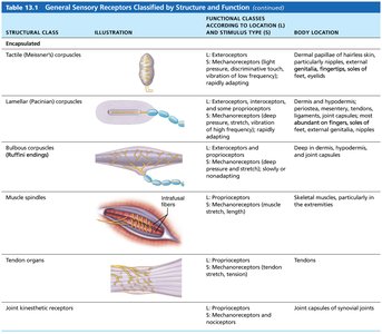
Simple Receptors: Encapsulated
Tactile (Meissner's) corpuscles
Lamellar (Pacinian) corpuscles
Bulbous corpuscles (Ruffini endings)
Muscle spindles
Tendon organs
Joint kinesthetic receptors
Sensation and Perception
From Sensation to Perception
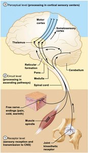
Sensation is the awareness of changes in the internal and external environment, while perception is the conscious interpretation of those stimuli. Survival depends on both processes.
Three levels of neural integration:
Receptor level – sensory receptors
Circuit level – ascending pathways
Perceptual level – neuronal circuits in the cerebral cortex
Pain and Referred Pain
Nociception: Perception of Pain
Nociceptors warn of actual or impending tissue damage, prompting protective actions. Pain stimuli include extreme pressure/temperature, histamine, K+, ATP, acids, and bradykinin. Pain impulses travel on fibers releasing neurotransmitters (glutamate, substance P), and can be blocked by endogenous opioids (e.g., endorphins).
Pain threshold: All perceive pain at the same stimulus intensity.
Pain tolerance: Varies among individuals; "sensitive to pain" means low tolerance, not low threshold.
Visceral and Referred Pain
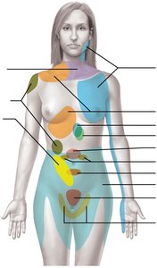
Visceral pain arises from stimulation of visceral organ receptors, felt as vague aching, gnawing, or burning. Referred pain is pain from one region perceived as coming from another, due to shared nerve pathways (e.g., left arm pain during heart attack).
Structure of a Nerve
Nerve Anatomy
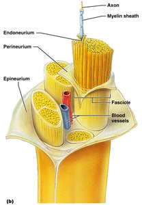
A nerve is a cordlike organ in the PNS, consisting of bundles of myelinated and nonmyelinated axons enclosed by connective tissue. Nerves are classified as spinal or cranial, based on their origin.
Endoneurium: Surrounds individual axons.
Perineurium: Bundles fibers into fascicles.
Epineurium: Encloses the entire nerve.
Classification of Nerves
Functional Classification
Sensory (afferent) nerves: Carry impulses to the CNS.
Motor (efferent) nerves: Carry impulses from the CNS.
Mixed nerves: Contain both sensory and motor fibers; most nerves are mixed.
Mixed nerves carry somatic and autonomic (visceral) impulses. Ganglia are collections of neuron cell bodies associated with nerves in the PNS.
Cranial Nerves
Overview of Cranial Nerves
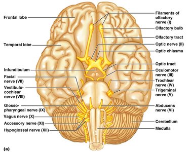
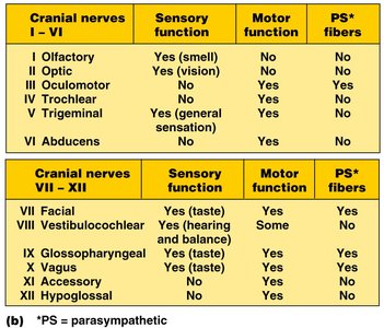
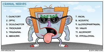
There are twelve pairs of cranial nerves, each with sensory, motor, or both functions. Four cranial nerves carry parasympathetic fibers. Each nerve is identified by a Roman numeral and a name.
Summary of Cranial Nerve Functions
Cranial Nerve | Sensory Function | Motor Function | Parasympathetic Fibers |
|---|---|---|---|
I Olfactory | Yes (smell) | No | No |
II Optic | Yes (vision) | No | No |
III Oculomotor | No | Yes | Yes |
IV Trochlear | No | Yes | No |
V Trigeminal | Yes (general sensation) | Yes | No |
VI Abducens | No | Yes | No |
VII Facial | Yes (taste) | Yes | Yes |
VIII Vestibulocochlear | Yes (hearing and balance) | No | No |
IX Glossopharyngeal | Yes (taste) | Yes | Yes |
X Vagus | Yes (taste) | Yes | Yes |
XI Accessory | No | Yes | No |
XII Hypoglossal | No | Yes | No |
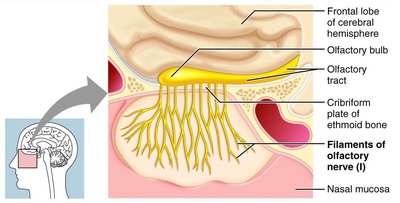
Cranial Nerve I: Olfactory
Arises from the olfactory epithelium, passes through the cribriform plate, and terminates in the primary olfactory cortex. Solely responsible for the sense of smell.
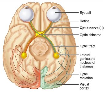
Cranial Nerve II: Optic
Originates from the retina, passes through optic canals, converges at the optic chiasm, synapses in the thalamus, and projects to the visual cortex. Solely responsible for vision.
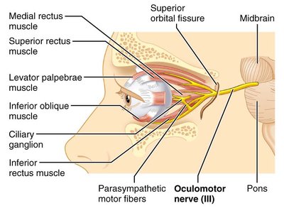
Cranial Nerve III: Oculomotor
Extends from the ventral midbrain to extrinsic eye muscles. Functions include raising eyelid, directing eyeball, constricting iris, and controlling lens shape. Parasympathetic cell bodies are in the ciliary ganglia.
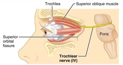
Cranial Nerve IV: Trochlear
Emerges from the dorsal midbrain, enters the orbit via the superior orbital fissure, and innervates the superior oblique muscle. Primarily a motor nerve directing the eyeball.
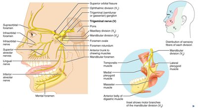
Cranial Nerve V: Trigeminal
Has three divisions: ophthalmic (V1), maxillary (V2), and mandibular (V3). Conveys sensory impulses from the face and supplies motor fibers for mastication.
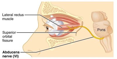
Cranial Nerve VI: Abducens
Leaves the inferior pons, enters the orbit via the superior orbital fissure, and innervates the lateral rectus muscle. Primarily a motor nerve.
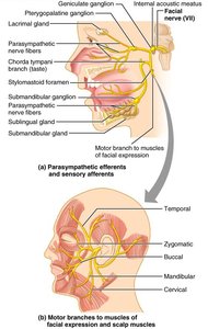
Cranial Nerve VII: Facial
Leaves the pons, travels through the internal acoustic meatus, and emerges on the lateral aspect of the face. Mixed nerve with motor functions (facial expression, lacrimal and salivary glands) and sensory function (taste from anterior two-thirds of tongue).
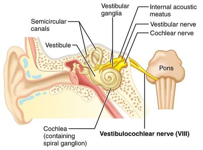
Cranial Nerve VIII: Vestibulocochlear
Arises from the hearing and equilibrium apparatus of the inner ear, passes through the internal acoustic meatus, and enters the brainstem. Two divisions: cochlear (hearing) and vestibular (balance).
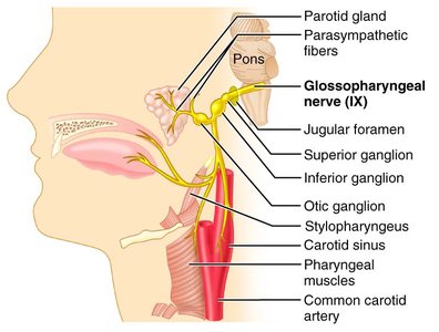
Cranial Nerve IX: Glossopharyngeal
Emerges from the medulla, leaves the skull via the jugular foramen, and runs to the throat. Mixed nerve with motor (tongue, pharynx, parotid gland) and sensory (taste, general sensation) functions.
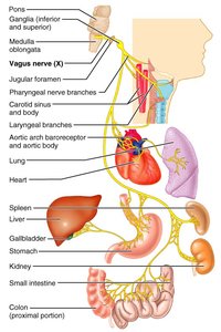
Cranial Nerve X: Vagus
The only cranial nerve extending beyond the head and neck. Mixed nerve with motor fibers (parasympathetic to heart, lungs, visceral organs) and sensory function (taste).
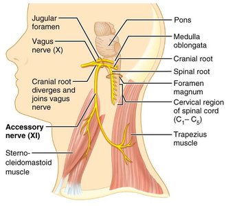
Cranial Nerve XI: Accessory
Formed from cranial and spinal roots, supplies fibers to the larynx, pharynx, soft palate, trapezius, and sternocleidomastoid muscles.
Cranial Nerve XII: Hypoglossal
Arises from the medulla, exits via the hypoglossal canal, and innervates muscles of the tongue for swallowing and speech.
Spinal Nerves
Overview of Spinal Nerves
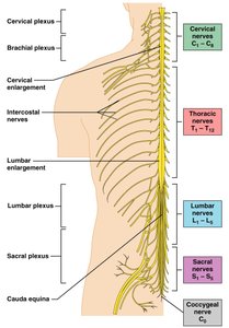
There are 31 pairs of mixed spinal nerves arising from the spinal cord, supplying all parts of the body except the head. They are named according to their point of issue: 8 cervical, 12 thoracic, 5 lumbar, 5 sacral, and 1 coccygeal.
Nerve Plexuses
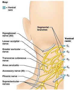
Ventral rami (except T2-T12) form interlacing nerve networks called plexuses in the cervical, brachial, lumbar, and sacral regions. Each branch contains fibers from several spinal nerves, ensuring redundancy in muscle innervation.
Dermatomes
Dermatome Mapping
A dermatome is the area of skin innervated by the cutaneous branches of a single spinal nerve. All spinal nerves except C1 participate in dermatomes. Most dermatomes overlap, so destruction of a single spinal nerve does not cause complete numbness.
Reflexes and Reflex Arcs
Reflexes
A reflex is a rapid, predictable motor response to a stimulus. Reflexes may be inborn (intrinsic) or learned (acquired), and can involve only peripheral nerves and the spinal cord or higher brain centers.
Reflex Arc Components
Receptor: Site of stimulus action
Sensory neuron: Transmits afferent impulses to CNS
Integration center: Monosynaptic or polysynaptic region within CNS
Motor neuron: Conducts efferent impulses to effector organ
Effector: Muscle fiber or gland responding to efferent impulse
Reflex Classification
Somatic reflexes: Activate skeletal muscle
Autonomic (visceral) reflexes: Activate visceral effectors (smooth/cardiac muscle or glands)
Muscle Spindles and Tendon Organs
Muscle Spindles
Muscle spindles are composed of intrafusal fibers, wrapped with sensory endings and innervated by gamma (γ) efferent fibers. Contractile muscle fibers (extrafusal) are innervated by alpha (α) efferent fibers.
Operation of Muscle Spindles
Stretching the muscle activates the spindle, increasing action potential rate in Ia fibers.
Contracting the muscle reduces tension, decreasing action potential rate.
α-γ coactivation maintains spindle tension and sensitivity during contraction.
Stretch Reflex
Stretched muscle spindles initiate a stretch reflex, causing contraction of the stretched muscle and inhibition of its antagonist (e.g., patellar reflex).
Tendon Reflex
The tendon reflex is the opposite of the stretch reflex, preventing damage due to excessive stretch. Contracting the muscle activates Golgi tendon organs, leading to relaxation of the contracting muscle and activation of the antagonist.
Flexor and Crossed Extensor Reflexes
The flexor (withdrawal) reflex is initiated by a painful stimulus, causing automatic withdrawal. The crossed extensor reflex involves withdrawal on the stimulated side and extension on the contralateral side, important for survival.
Superficial Reflexes
Plantar reflex: Tests integrity of cord from L4 to S2; abnormal response is Babinski's sign.
Abdominal reflex: Tests integrity of cord from T8 to T12; response is contraction of abdominal muscles and movement of umbilicus toward stimulus.
References: Marieb, E. N., & Hoehn, K. (2023). Human anatomy and physiology (12th ed.). Pearson.
