 Back
BackPeripheral Nervous System and Reflex Activity: Study Guide
Study Guide - Smart Notes
 Tailored notes based on your materials, expanded with key definitions, examples, and context.
The Peripheral Nervous System (PNS)
Overview and Structural Organization
The Peripheral Nervous System (PNS) serves as the communication link between the Central Nervous System (CNS) and the rest of the body. It consists of all neural structures outside the brain and spinal cord, including sensory receptors, peripheral nerves, ganglia, and motor endings. The PNS allows the CNS to receive information and make decisions, facilitating both sensory input and motor output.
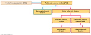
Sensory Receptors: Detect changes in the environment.
Transmission Lines: Peripheral nerves and ganglia transmit signals.
Motor Endings: Activate effectors such as muscles and glands.
Somatosensory System and Sensory Receptors
General Organization
The somatosensory system is responsible for processing sensory information from the body wall and limbs. It relies on sensory receptors to detect stimuli, which must excite a receptor and generate an action potential (AP) to reach the cortex for sensation and perception.
Sensation: Awareness of a stimulus, both internal and external.
Perception: Interpretation of the meaning of a stimulus.
Classification by Stimulus Type
Mechanoreceptors: Respond to touch, pressure, vibration, and stretch.
Thermoreceptors: Sensitive to temperature changes.
Photoreceptors: Respond to light energy (e.g., retina).
Chemoreceptors: Respond to chemicals (e.g., smell, taste, blood chemistry).
Nociceptors: Sensitive to pain-causing stimuli.
Classification by Location
Exteroceptors: Detect stimuli from outside the body (touch, pressure, pain, temperature).
Interoceptors: Detect stimuli from internal viscera and blood vessels (chemical changes, tissue stretch, temperature).
Proprioceptors: Detect internal stimuli in muscles, tendons, joints, ligaments, and connective tissue; inform the brain of body movements.
Transmission Lines: Nerves and Ganglia
Structure and Classification of Nerves
Nerves are cordlike organs in the PNS, classified as cranial or spinal. Each nerve is a bundle of peripheral axons. Collections of neuron cell bodies are called nuclei in the CNS and ganglia in the PNS.
Mixed Nerves: Contain both sensory and motor fibers; impulses travel to and from the CNS.
Sensory (Afferent) Nerves: Carry impulses toward the CNS.
Motor (Efferent) Nerves: Carry impulses away from the CNS.
Most nerves are mixed, including somatic and visceral afferent/efferent fibers. Ganglia associated with afferent fibers are dorsal root ganglia (sensory, somatic), while those with efferent fibers are autonomic ganglia (motor, visceral).
Cranial and Spinal Nerves
Cranial Nerves
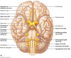
There are 12 pairs of cranial nerves associated with the brain. Most are mixed nerves, but two pairs are purely sensory. Cranial nerves are essential for sensory and motor functions of the head and neck.
Spinal Nerves
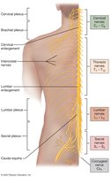
There are 31 pairs of spinal nerves, each containing thousands of fibers. They arise from the spinal cord and supply all body parts except the head and part of the neck. All are mixed nerves, named for their point of issue from the spinal cord.
8 pairs of cervical nerves (C1–C8)
12 pairs of thoracic nerves (T1–T12)
5 pairs of lumbar nerves (L1–L5)
5 pairs of sacral nerves (S1–S5)
1 pair of coccygeal nerves (C0)
Spinal Nerve Structure
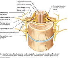
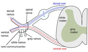
Each spinal nerve connects to the spinal cord via two roots:
Ventral Roots: Contain motor (efferent) fibers; innervate skeletal muscles.
Dorsal Roots: Contain sensory (afferent) fibers; conduct impulses from peripheral receptors.
Ventral and dorsal roots branch medially as rootlets and join laterally to form the spinal nerve.
Spinal Nerve Branches and Rami
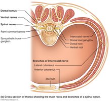
Spinal nerves emerge from the vertebral column via intervertebral foramina and immediately divide into three branches:
Dorsal Ramus: Smaller branch; supplies posterior body trunk.
Ventral Ramus: Larger branch; supplies the rest of the trunk and limbs.
Meningeal Branch: Reenters vertebral canal to innervate meninges and blood vessels.
Rami Communicantes: Contain autonomic nerve fibers; join ventral rami in the thoracic region.
Roots vs. Rami
Roots: Medial to and form spinal nerves; each root is purely sensory or motor.
Rami: Distal to and are lateral branches of spinal nerves; can carry both sensory and motor fibers.
Nerve Plexuses
Formation and Function
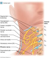
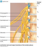
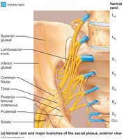
Ventral rami form interlacing nerve networks called nerve plexuses in the cervical, brachial, lumbar, and sacral regions. Within a plexus, fibers crisscross so that each branch contains fibers from several different spinal nerves. This arrangement ensures that each limb muscle is innervated by more than one spinal nerve, so damage to one does not cause paralysis.
Cervical Plexus
The cervical plexus is formed by ventral rami of C1–C4 and supplies nerves to the neck and upper shoulder.
Lumbar Plexus
The lumbar plexus is formed by ventral rami of L1–L4 and supplies nerves to the lower abdomen, thigh, and groin.
Sacral Plexus
The sacral plexus is formed by ventral rami of L4–S4 and supplies nerves to the pelvis, buttocks, and lower limbs.
Reflex Activity
Reflex Arc and Types of Reflexes
Reflexes are rapid, involuntary, and predictable motor responses to stimuli. They can be inborn (intrinsic) or learned (acquired). Reflexes help maintain posture and control visceral activities, and can be modified by learning and conscious effort.
Inborn Reflexes: Automatic responses to stimuli.
Learned Reflexes: Acquired through practice or repetition.
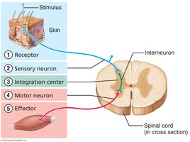
Components of a Reflex Arc
A reflex arc consists of five basic components:
Receptor: Site of stimulus action.
Sensory Neuron: Transmits afferent impulses to the CNS.
Integration Center: Monosynaptic or polysynaptic region within the CNS.
Motor Neuron: Conducts efferent impulses from the integration center to the effector organ.
Effector: Muscle fiber or gland cell that responds to efferent impulses.
Classification of Reflexes
Somatic Reflexes: Activate skeletal muscle.
Autonomic (Visceral) Reflexes: Activate visceral effectors (smooth or cardiac muscle, glands).
Spinal reflexes occur without direct involvement of higher brain centers, but the brain is still advised of activity and may influence the reflex.
Flexor and Crossed-Extensor Reflexes
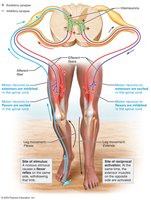
The flexor (withdrawal) reflex is initiated by a painful stimulus and causes automatic withdrawal of the threatened body part. It is ipsilateral and polysynaptic, involving multiple muscles. The crossed-extensor reflex occurs with flexor reflexes in weight-bearing limbs to maintain balance, consisting of ipsilateral withdrawal and contralateral extension.
Flexor Reflex: Protective, important for survival; can be overridden by the brain.
Crossed-Extensor Reflex: Maintains balance during withdrawal; example: stepping on broken glass.
Summary Table: Classification of Sensory Receptors
Type | Stimulus Detected | Location |
|---|---|---|
Mechanoreceptor | Touch, pressure, vibration, stretch | Skin, muscles |
Thermoreceptor | Temperature changes | Skin, internal organs |
Photoreceptor | Light | Retina |
Chemoreceptor | Chemicals | Nose, tongue, blood vessels |
Nociceptor | Pain | Skin, internal organs |
Summary Table: Spinal Nerve Classification
Type | Direction | Function |
|---|---|---|
Mixed | To and from CNS | Sensory and motor |
Sensory (Afferent) | Toward CNS | Sensory only |
Motor (Efferent) | Away from CNS | Motor only |
Key Equations
Action Potential Generation:
The generation of an action potential (AP) in a neuron can be described by the change in membrane potential:
Where is the change in membrane potential, is the final membrane potential after stimulus, and is the resting membrane potential.
Nerve Impulse Transmission:
The speed of nerve impulse transmission depends on axon diameter and myelination:
Where is the velocity, is the distance traveled, and is the time taken.
Additional info: Academic context was added to clarify the structure and function of nerves, plexuses, and reflexes, as well as to provide summary tables and equations for exam preparation.
