 Back
BackPeripheral Nervous System and Reflexes: Structure, Function, and Clinical Relevance
Study Guide - Smart Notes
 Tailored notes based on your materials, expanded with key definitions, examples, and context.
Peripheral Nervous System and Reflexes
Overview of the Nervous System
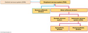
The nervous system is divided into the central nervous system (CNS) and the peripheral nervous system (PNS). The CNS consists of the brain and spinal cord, while the PNS includes all neural structures outside the CNS. The PNS is further subdivided into sensory (afferent) and motor (efferent) divisions, with the motor division containing both the somatic and autonomic nervous systems.
Major Functions of the Nervous System
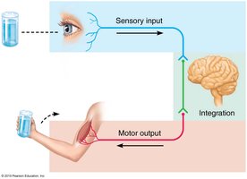
Sensation: Detection of changes in the internal and external environment, carried by afferent nerves of the PNS. Sensations include touch, pressure, vibration, temperature, pain, proprioception, and special senses (sight, hearing, taste, smell, equilibrium).
Integration: Processing and interpretation of sensory input, occurring in the CNS.
Response: Activation of effector organs (muscles and glands) via efferent nerves of the PNS to produce a response.
Sensory Receptors and Signal Processing
Classification of Sensory Receptors
Sensory receptors are specialized to transduce environmental stimuli into action potentials. They can be classified by:
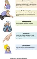
Type of Stimulus:
Thermoreceptors: Detect temperature changes.
Nociceptors: Detect pain.
Mechanoreceptors: Detect pressure, vibration, and stretch.
Photoreceptors: Detect light (vision).
Chemoreceptors: Detect chemical changes (taste, smell, blood chemistry).
Osmoreceptors: Detect solute concentrations.
Location:
Exteroceptors: Sense external environment (e.g., touch, taste).
Interoceptors: Sense internal environment (e.g., organ distension, chemical changes).
Proprioceptors: Sense body position and movement (found in muscles and joints).
Cell Type:
Free nerve endings
Encapsulated endings
Specialized receptor cells
Sensory Signal Transmission and Processing
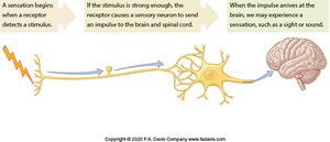
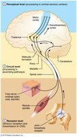
Sensory information is transmitted through a series of steps:
Level 1: Sensory Receptors – Stimulus must reach threshold to generate an action potential. Sensory adaptation can occur, reducing sensitivity to constant stimuli (e.g., touch receptors adapt quickly, nociceptors do not).
Level 2: Circuit Level – Processing in ascending pathways, typically involving three neurons (first-order, second-order, third-order). Pathways include dorsal columns (touch, proprioception), spinothalamic tracts (pain, temperature), and spinocerebellar tracts (subconscious proprioception).
Level 3: Perceptual Level – Sensory information is interpreted in the cerebral cortex, allowing for magnitude estimation, spatial discrimination, quality discrimination, and pattern recognition.
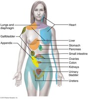
Referred Pain
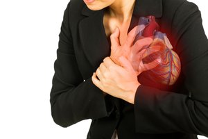
Visceral pain afferents travel the same pathways as somatic pain afferents, leading to referred pain—pain perceived at a location other than the site of the painful stimulus. For example, heart attack pain may be felt in the left arm or jaw.
Nerves and Peripheral Organization
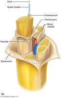
Structure of a Nerve
A nerve is a bundle of axons in the PNS, organized into fascicles and surrounded by connective tissue layers: endoneurium (around each axon), perineurium (around each fascicle), and epineurium (around the entire nerve). Nerves contain both myelinated and unmyelinated fibers, as well as blood and lymphatic vessels.
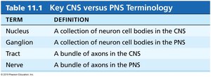
Key CNS vs PNS Terminology
Term | Definition |
|---|---|
Nucleus | A collection of neuron cell bodies in the CNS |
Ganglion | A collection of neuron cell bodies in the PNS |
Tract | A bundle of axons in the CNS |
Nerve | A bundle of axons in the PNS |
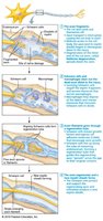
Regeneration of Nerves
CNS axons: Generally cannot regenerate due to inhibitory factors from oligodendrocytes and scar formation by astrocytes.
PNS axons: Can regenerate if the cell body is intact and the cut ends are aligned. Schwann cells form a regeneration tube to guide axonal regrowth, but the process is slow and requires retraining.
Cranial and Spinal Nerves
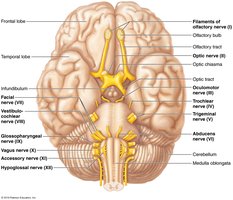
Twelve Pairs of Cranial Nerves
There are 12 pairs of cranial nerves, each with specific sensory, motor, or mixed functions. They originate from the brain or brainstem and innervate structures primarily in the head and neck.
I – Olfactory (Sensory): Smell
II – Optic (Sensory): Vision
III – Oculomotor (Motor): Eye movement, pupil constriction
IV – Trochlear (Motor): Eye movement
V – Trigeminal (Both): Facial sensation, mastication
VI – Abducens (Motor): Eye movement
VII – Facial (Both): Taste (anterior 2/3 tongue), facial expression, glands
VIII – Vestibulocochlear (Sensory): Hearing, equilibrium
IX – Glossopharyngeal (Both): Taste (posterior 1/3 tongue), pharynx, carotid body
X – Vagus (Both): Parasympathetic control of thoracic/abdominal organs, taste from epiglottis
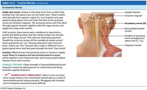
XI – Accessory (Motor): Sternocleidomastoid, trapezius
XII – Hypoglossal (Motor): Tongue muscles
Spinal Nerves and Plexuses
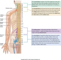
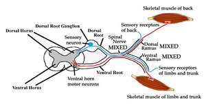
There are 31 pairs of spinal nerves, named for their region of emergence: 8 cervical, 12 thoracic, 5 lumbar, 5 sacral, and 1 coccygeal. Each spinal nerve is formed by the joining of dorsal (sensory) and ventral (motor) roots and quickly branches into dorsal and ventral rami. The ventral rami form plexuses (cervical, brachial, lumbar, sacral) that innervate the limbs and trunk.
Dermatomes and Peripheral Nerve Maps
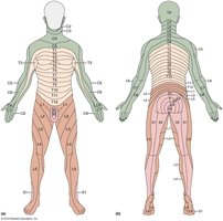
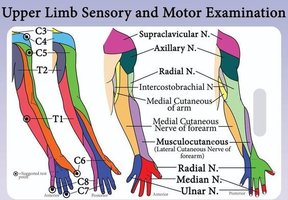
A dermatome is an area of skin innervated by a single spinal nerve. Peripheral nerve maps show the cutaneous distribution of named peripheral nerves, which may include fibers from multiple spinal levels. Knowledge of dermatomes is essential for neurological assessment.
Motor Control and Reflexes
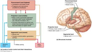
Levels of Motor Control
Motor control is organized into three hierarchical levels:
Segmental Level: Spinal cord circuits (reflexes and central pattern generators).
Projection Level: Motor cortex and brainstem nuclei, which convey instructions to the spinal cord.
Precommand Level: Cerebellum and basal nuclei, which regulate and plan movements.
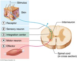
The Reflex Arc
A reflex arc is the basic functional unit of the nervous system, allowing for rapid, automatic responses to stimuli. It consists of five components:
Receptor
Sensory neuron
Integration center
Motor neuron
Effector
Types of Reflexes
Intrinsic (Inborn) Reflexes: Rapid, predictable motor responses (e.g., knee-jerk, withdrawal from pain).
Learned (Acquired) Reflexes: Result from practice or repetition (e.g., riding a bike, driving).


Somatic and Autonomic Reflexes
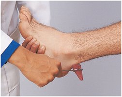
Somatic Reflexes: Involve skeletal muscles. Examples include stretch reflexes (patellar, calcaneal, biceps), superficial reflexes (corneal, gag), and the plantar reflex (Babinski sign).
Autonomic (Visceral) Reflexes: Involve smooth muscle, cardiac muscle, or glands. Examples include pupillary light reflex, salivary reflex, and reflexes controlling digestion, sweating, and blood pressure.

Clinical Relevance of Reflexes
Hypoactive reflexes: May indicate lower motor neuron injury.
Hyperactive reflexes: May indicate upper motor neuron injury.
Referred pain: Important for differential diagnosis in clinical practice.
Special Reflexes
Flexor Withdrawal Reflex: Rapid withdrawal from a painful stimulus.
Crossed Extensor Reflex: Maintains balance when a limb is withdrawn.
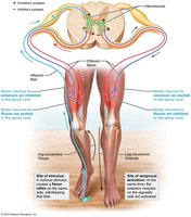
Additional info: Reflexes are essential for homeostasis and survival, providing rapid responses to potentially harmful stimuli and maintaining posture and vital functions without conscious thought.
